Home > Science > SEM
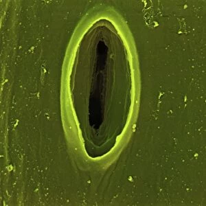
Dividing yeast cells, SEM

![]()
Wall Art and Photo Gifts from Science Photo Library
Dividing yeast cells, SEM
Dividing yeast cells. Coloured scanning electron micrograph (SEM) of Schizosaccharomyces pombe yeast cells dividing. S. pombe is a single-celled fungus that is studied widely as a model organism for eukaryotic cell division. It is a rod-shaped yeast that grows by elongation at its ends. It replicates by binary fission. When it reaches a certain size its genetic material (deoxyribonucleic acid, DNA) separates to opposite ends of the cell and a division septum (wall) grows across the centre of the cell, dividing it into two daughter cells that are identical to the parent cell. Magnification: x5500 when printed 10cm wide
Science Photo Library features Science and Medical images including photos and illustrations
Media ID 6291947
© STEVE GSCHMEISSNER/SCIENCE PHOTO LIBRARY
Asexual Binary Fission Dividing Division Eukaryote Eukaryotic Eumycota Fungal Fungi Fungus Model Organism Mycology Naturemycology Re Production Replicating Replication Reproducing Single Celled Yeast Cells False Coloured Micro Biology Microbiological
EDITORS COMMENTS
In this coloured scanning electron micrograph (SEM), we witness the intricate process of cell division in Schizosaccharomyces pombe, a single-celled fungus renowned as a widely studied model organism for eukaryotic cell division. The rod-shaped yeast, S. pombe, grows by elongation at its ends and replicates through binary fission. As the yeast cell reaches a certain size, its genetic material (DNA) begins to separate to opposite ends of the cell. The division septum, a thin wall, then grows across the centre of the cell, dividing it into two identical daughter cells. This process, known as medial fission, is a hallmark of S. pombe and is a crucial aspect of its asexual replication. This stunning SEM image, with a magnification of x5500 when printed 10cm wide, offers a mesmerizing glimpse into the microscopic world of biology. The false colours enhance the visualization of the intricate details of the dividing yeast cells, revealing the complex structures involved in the process of cell division. S. pombe is a significant organism in various fields of study, including microbiology, mycology, and eukaryote biology. Its unique mode of binary fission and the ease with which it can be cultured and manipulated make it an invaluable tool for researchers investigating the fundamental mechanisms of cell division in eukaryotes. This image serves as a testament to the beauty and complexity of nature, showcasing the intricacies of cellular processes that underpin all life on Earth.
MADE IN THE USA
Safe Shipping with 30 Day Money Back Guarantee
FREE PERSONALISATION*
We are proud to offer a range of customisation features including Personalised Captions, Color Filters and Picture Zoom Tools
SECURE PAYMENTS
We happily accept a wide range of payment options so you can pay for the things you need in the way that is most convenient for you
* Options may vary by product and licensing agreement. Zoomed Pictures can be adjusted in the Cart.