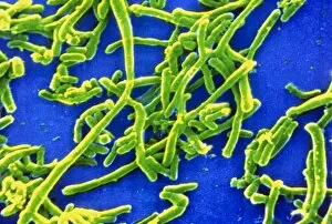
Bacteria Collection (#37)

"Bacteria: The Microscopic Marvels of Yellowstone National Park" In the heart of Wyoming, USA lies the breathtaking Yellowstone National Park

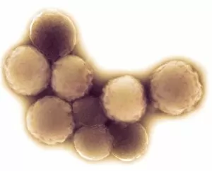
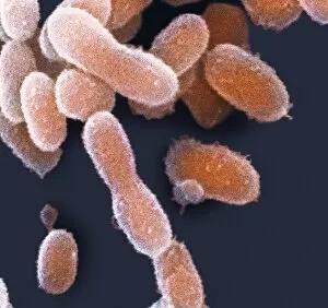
Background imageBacteria Collection: Cold-loving extremophile bacteria, SEM

Cold-loving extremophile bacteria, SEM
Cold-loving extremophile bacteria. Coloured scanning electron micrograph (SEM) of Rhodoglobus vestalii. This psychrophilic (cold-loving) bacteria was retrieved from a lake in Antarctica

Background imageBacteria Collection: Geobacter metallireducens bacterium, TEM

Geobacter metallireducens bacterium, TEM
Geobacter metallireducens bacterium, coloured transmission electron micrograph. This anaerobic bacteriaoxidises organic compounds to form carbon dioxide

Background imageBacteria Collection: F / col TEM of Enterococcus faecalis

F / col TEM of Enterococcus faecalis
Enterococcus faecalis. Coloured transmission electron micrograph (TEM) of Enterococcus faecalis bacteria (previously known as Streptococcus faecalis)

Background imageBacteria Collection: Streptococcus mutans bacteria on tooth

Streptococcus mutans bacteria on tooth
Streptococcus mutans bacteria. Coloured scanning electron micrograph (SEM) of Streptococcus mutans bacteria adhering to the enamel of a tooth

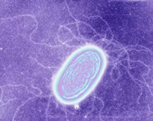
Background imageBacteria Collection: Geobacter metallireducens bacteria, TEM

Geobacter metallireducens bacteria, TEM
Geobacter metallireducens bacterium, coloured transmission electron micrograph. This anaerobic bacteriaoxidises organic compounds to form carbon dioxide

Background imageBacteria Collection: Resistant Streptococcus pyogenes strain

Resistant Streptococcus pyogenes strain
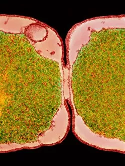
Resistant Streptococcus strain. Coloured transmission electron micrograph of a dividing Streptococcus pyogenes group A bacterium

Background imageBacteria Collection: Mycoplasma mycoides bacteria

Mycoplasma mycoides bacteria
Mycoplasma mycoides, coloured transmission electron micrograph (TEM). M. mycoides has a very small genome (total genetic material)

Background imageBacteria Collection: Nitrogen-fixing bacteria, SEM

Nitrogen-fixing bacteria, SEM
Nitrogen-fixing bacteria. Coloured scanning electron micrograph (SEM) of nitrogen-fixing soil bacteria (Rhizobium sp.) living on a root nodule of a runner bean plant (Phaseolus coccineus)

Background imageBacteria Collection: Root nodules of white clover

Root nodules of white clover

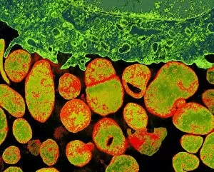
Background imageBacteria Collection: Mycoplasma bacteria, TEM

Mycoplasma bacteria, TEM
Mycoplasma bacteria. Coloured transmission electron micrograph (TEM) of a section through a group of Mycoplasma sp. bacteria (orange) surrounding an infected cell (green)

Background imageBacteria Collection: Coloured SEM of streptococcus pneumoniae

Coloured SEM of streptococcus pneumoniae
False colour scanning electron micrograph (SEM) of Streptococcus pneumoniae (pneumonococcus), a spherical, Gram-positive bacterium associated with pneumonia in humans

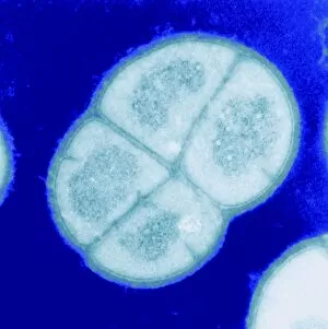
Background imageBacteria Collection: Radiation resistant bacteria, SEM

Radiation resistant bacteria, SEM
Radiation resistant bacteria. Coloured scanning electron micrograph (SEM) of four Deinococcus radiodurans bacteria forming a tetrad

Background imageBacteria Collection: Root nodule of pea plant

Root nodule of pea plant

Background imageBacteria Collection: Mycoplasma genitalium bacteria

Mycoplasma genitalium bacteria
Mycoplasma genitalium, coloured transmission electron micrograph (TEM). M. genitalium has the smallest genome (total genetic material) of all living organisms

Background imageBacteria Collection: Root nodule of white clover plant

Root nodule of white clover plant
False-colour scanning electron micrograph of a root nodule on the white clover plant, Trifolium repens, caused by the nitrogen-fixing bacteria Rhizobium trifolii

Background imageBacteria Collection: Archaeoglobus bacteria

Archaeoglobus bacteria
Thermophilic bacteria. False colour light micrograph of the thermophilic bacteria Archaeoglobus fulgidus. This organism occurs in hot sediments near submarine hydrothermal vents

Background imageBacteria Collection: Ultra-small extremophile bacteria, SEM

Ultra-small extremophile bacteria, SEM
Ultra-small extremophile bacteria. Coloured scanning electron micrograph (SEM) of Chryseobacterium greenlandensis. This bacteria was retrieved from a 120
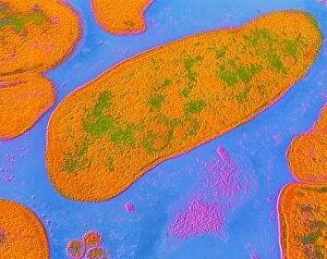
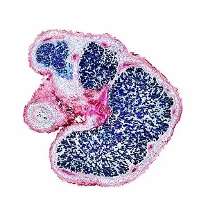
Background imageBacteria Collection: Legume root nodules, light micrograph

Legume root nodules, light micrograph
Legume root nodules. Light micrograph of sections through two root nodules from a broad bean plant (Vicia faber). These root nodules are full of nitrogen-fixing bacteria

Background imageBacteria Collection: Broad bean root nodules, light micrograph

Broad bean root nodules, light micrograph
Broad bean root nodules. Light micrograph of the sections of the root nodules of the broad bean (Vicia faber) which are full of nitrogen-fixing bacteria

Background imageBacteria Collection: Gut bacterium reproducing, TEM

Gut bacterium reproducing, TEM
Gut bacterium reproducing. Coloured transmission electron micrograph (TEM) of a single bacterium dividing into two identical clone daughter bacteria

Background imageBacteria Collection: Spread of disease

Spread of disease. Conceptual computer artwork of Earth over-run with bacteria

Background imageBacteria Collection: Branhamella catarrhalis bacteria

Branhamella catarrhalis bacteria
False-colour transmission electron micrograph (TEM) of Branhamella catarrhalis, formerly known as Neisseria catarrhalis, a species of Gram- negative bacteria which exists as one of the commonest

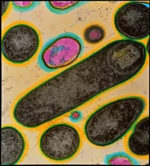
Background imageBacteria Collection: Clostridium bacteria

Clostridium bacteria
Coloured transmission electron micrograph (TEM) of Clostridium botulinum bacteria, the cause of botulism food poisoning. The bacterium seen in longitudinal section at centre is in the process of

Background imageBacteria Collection: Coloured TEM of E. coli bacteria dividing

Coloured TEM of E. coli bacteria dividing
E. Coli bacteria dividing. Transmission electron micrograph of the division zone between two new Escherichia coli bacteria showing the cells undergoing binary fission

Background imageBacteria Collection: Colony of E. coli bacteria

Colony of E. coli bacteria
False-colour scanning electron micrograph of a colony of the rod-shaped, Gram-negative bacterium, Escherichia coli. E. coli, as it is commonly known, is found in the human intestines

Background imageBacteria Collection: Bacteria on skin

Bacteria on skin. Computer artwork of bacteria (blue and green) on human skin. Many types of bacteria are found on human skin, especially associated with sweat glands and hair follicles

Background imageBacteria Collection: Plague bacteria

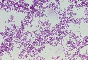
Plague bacteria. Light micrograph of Gram- stained Yersinia pestis bacteria. This bacterium is the cause of bubonic plague (generally agreed to have been the Black Death of the Middle Ages)

Background imageBacteria Collection: Legionella pneumophila bacteria

Legionella pneumophila bacteria

Background imageBacteria Collection: Bacteria, computer artwork

Bacteria, computer artwork
Bacteria. Computer artwork of a multitude of rod-shaped (bacillus) bacteria. Many bacteria have this shape, including Escherichia coli, which can cause food poisoning, and Bacillus anthracis

Background imageBacteria Collection: Clostridium botulinum bacteria with spores

Clostridium botulinum bacteria with spores
Coloured transmission electron micrograph (TEM) of the bacteria Clostridium botulinum, cause of botulism food poisoning in humans, in the process of sporulation (spore formation)

Background imageBacteria Collection: Bacteria in the nose, SEM

Bacteria in the nose, SEM
Bacteria in the nose. Coloured scanning electron micrograph (SEM) of bacteria (red) on the surface of the nasal cavity. One of the squamous epithelium cells (lower centre to lower right)

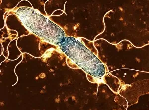
Background imageBacteria Collection: Salmonella, TEM

Salmonella, TEM
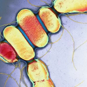
Salmonella. Coloured transmission electron micrograph (TEM) of Salmonella paratyphi B bacteria (also called Salmonella schotmulleri)

Background imageBacteria Collection: F / col TEM of Haemophilus influenzae

F / col TEM of Haemophilus influenzae
False-colour transmission electron micrograph (TEM) of Haemophilus influenzae (Pfeiffers bacillus), a Gram negative, non-motile, non- sporing species of bacilli (rod-shaped bacteria). H

Background imageBacteria Collection: Pseudomonas aeruginosa bacteria

Pseudomonas aeruginosa bacteria
False colour transmission electron micrograph of the rodlike bacteria Pseudomonas aeruginosa (gram negative). These bacteria are common in the soil, decomposing organic material & water

Background imageBacteria Collection: Colour SEM of Corynebacterium diphtheria

Colour SEM of Corynebacterium diphtheria
False-colour scanning electron micrograph (SEM) of Corynebacterium diphtheria, the Gram-positive, non-motile bacteria responsible for diphtheria

Background imageBacteria Collection: S. maltophilia bacteria, TEM

S. maltophilia bacteria, TEM
Stenotrophomonas maltophilia bacteria, coloured transmission electron micrograph (TEM). This aerobic Gram-negative bacterium, previously known as Pseudomonas maltophilia

Background imageBacteria Collection: Acinetobacter sp. bacteria

Acinetobacter sp. bacteria
Coloured transmission electron micrograph (TEM) of a colony of Acinetobacter sp. a species of non- motile, non-sporing, Gram negative bacteria belonging to the Neisseriaceae group

Background imageBacteria Collection: Artwork of structure of a bacterium

Artwork of structure of a bacterium
Cutaway illustration of the internal structure of a typical bacterial cell. The bacterial cell wall (orange & brown) overlies the cytoplasmic membrane (white)

Background imageBacteria Collection: Clostridium tetani bacterium

Clostridium tetani bacterium
False-colour transmission electron micrograph (TEM) of the bacterium Clostridium tetani, a Gram- positive, spore-forming, anaerobic bacillus (rod-shaped bacterium) that causes tetanus

Background imageBacteria Collection: Germ, conceptual computer artwork

Germ, conceptual computer artwork


For sale as Licensed Images

Choose your image, Select your licence and Download the media

"Bacteria: The Microscopic Marvels of Yellowstone National Park" In the heart of Wyoming, USA lies the breathtaking Yellowstone National Park, a haven for nature enthusiasts and scientists alike. Amidst its natural wonders, one can find an extraordinary display of microbial life at the Grand Prismatic Spring in the Midway Geyser Basin. Picture No. 11675590 captures this mesmerizing sight, showcasing vibrant hues that seem to dance upon the water's surface. However, what may appear as an artistic masterpiece is actually a thriving community of bacteria. Zooming in closer with SEM C018 / 8596 reveals a neutrophil engulfing MRSA - Methicillin-resistant Staphylococcus aureus. This image serves as a reminder of how these tiny organisms can pose significant threats to human health. Another SEM image showcases E. Coli bacteria, reminding us that not all it can harmful; some play crucial roles in our digestive system and even aid scientific research. Moving back to Yellowstone's enchantment, we encounter Salmonella bacteria through another captivating SEM photograph. These microscopic creatures serve as a stark reminder that even within such serene landscapes, dangers lurk unseen. Coloured TEM imagery introduces us to Yersinia pestis bacteria – responsible for causing devastating outbreaks like the infamous Black Death throughout history. Such visuals emphasize their intricate structures and highlight why understanding them is vital for public health efforts. Venturing beyond real-life images into computer artwork brings forth a beta DNA segment surrounded by spheres—a representation of genetic material essential for bacterial survival and reproduction. Artistic depictions also shed light on various cell types found within these microorganisms' complex world—each playing unique roles necessary for their survival and proliferation. Historical diagrams reveal cultures of Anthrax—an infectious disease with severe consequences if left unchecked—underscoring humanity's ongoing battle against bacterial infections throughout time. Tuberculosis bacteria make their appearance too—a haunting reminder that despite medical advancements, infectious diseases continue to challenge us.