
Blood Vessel Collection (#2)
"Exploring the intricate network of blood vessels in our body: from brain to lungs, groin to face and scalp


For sale as Licensed Images
Choose your image, Select your licence and Download the media
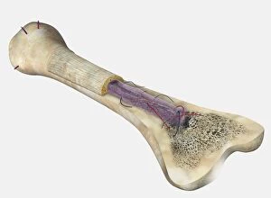
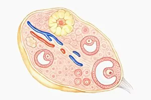
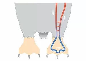
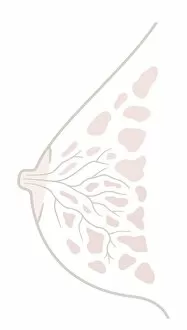
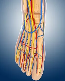
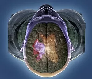
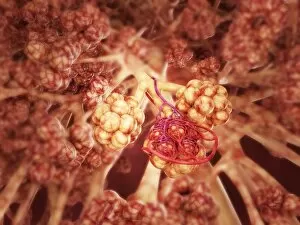
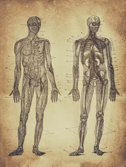
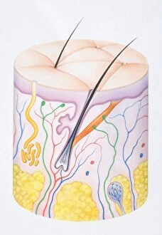
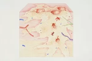
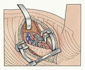
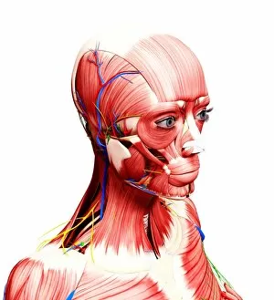
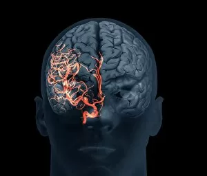
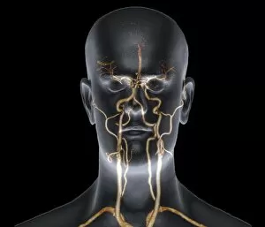
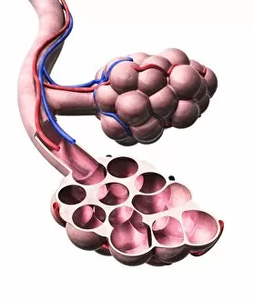
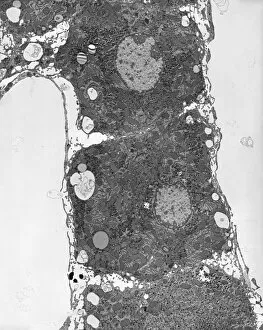
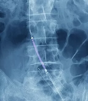
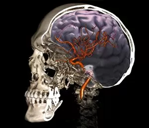
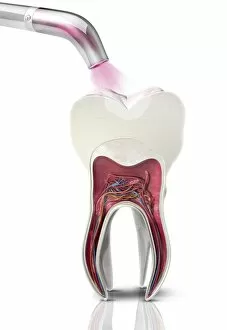
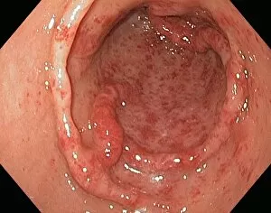
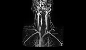
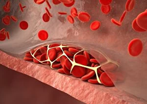
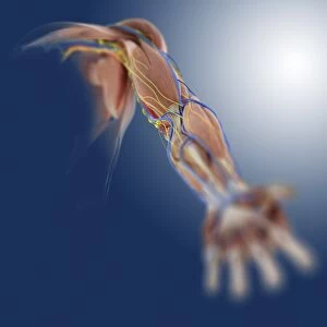
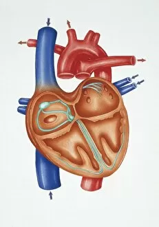
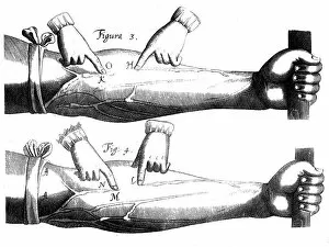
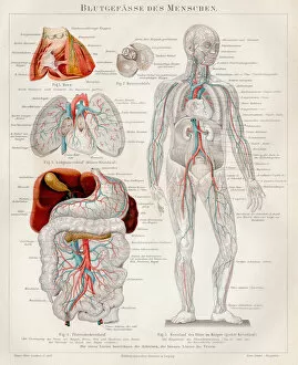
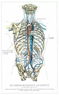
"Exploring the intricate network of blood vessels in our body: from brain to lungs, groin to face and scalp. " "Dive into the fascinating world of blood vessels with a 3D angiogram and a diagram showcasing their role in brain health. " "The vital connection between our lungs and bloodstream revealed through a detailed diagram highlighting the blood supply. " "Unveiling an artistic masterpiece from 1825, depicting male groin arteries - a testament to early anatomical studies. " "Discovering the superficial arteries and veins that grace our face and scalp, intricately illustrated for medical understanding. " "A mesmerizing artwork capturing the complexity of the blood coagulation cascade - an essential process for healing. " "Intriguing anatomy meets biology as we delve into colorful images showcasing various aspects function. " "Leonardo da Vinci's genius shines through pen and ink studies dating back to c1510, unraveling arm muscles alongside superficial vessels. " "With every beat, our human heart keeps us alive. Marvel at its beauty through captivating artwork that celebrates its strength. " "Journeying back in time with an exquisite 1825 artwork revealing head and chest arteries - a glimpse into historical medical knowledge. " "A close-up view of red blood cells captured by scanning electron microscopy showcases their unique structure within our bloodstream. " "Glimpsing at a healthy heart portrayed in stunning artwork; reminding us of this incredible organ's importance in maintaining life.