Blood Vessel Collection (#3)

"Exploring the intricate network of blood vessels in our body: from brain to lungs, groin to face and scalp

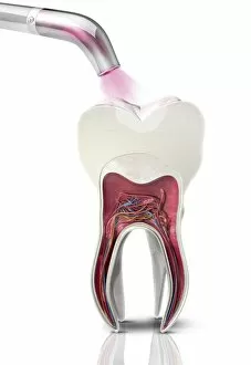
Background imageBlood Vessel Collection: Dental filling polymerisation, artwork C014 / 2025

Dental filling polymerisation, artwork C014 / 2025
Dental filling polymerisation. Artwork of a cross-section of a human tooth, with a filling (white) being set using an ultraviolet light lamp tool

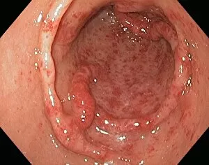
Background imageBlood Vessel Collection: Gastric antral vascular ectasia C016 / 8328

Gastric antral vascular ectasia C016 / 8328
Gastric antral vascular ectasia. Endoscopic view of the antrum (final portion of stomach), affected by gastric antral vascular ectasia (GAVE)

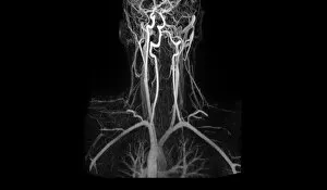
Background imageBlood Vessel Collection: Artery damage, MRA scan

Artery damage, MRA scan
Artery damage. Magnetic resonance angiography (MRA) scan of the neck of a 23-year-old male motorcycle crash victim. Dissection of the right internal carotid artery (left)

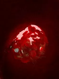
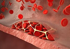
Background imageBlood Vessel Collection: Blood clot, artwork C016 / 4619

Blood clot, artwork C016 / 4619
Blood clot in a blood vessel, computer artwork. Red blood cells (erythrocytes) are trapped within a fibrin protein mesh (cream)

Background imageBlood Vessel Collection: Thrombosed blood vessel, artwork C013 / 4649

Thrombosed blood vessel, artwork C013 / 4649
Thrombosed blood vessel. Computer artwork of a blood clot (thrombus) formed on the internal wall of a blood vessel

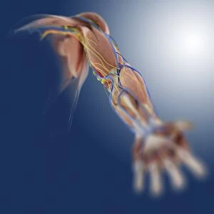
Background imageBlood Vessel Collection: Arm anatomy, artwork C013 / 4583

Arm anatomy, artwork C013 / 4583
Arm anatomy. Computer artwork of an oblique anterior view looking up a human arm, showing muscles and tendons (brown and white), bones and ligaments (white), arteries and veins (red and blue)

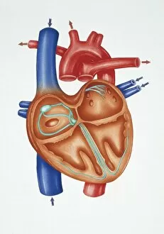
Background imageBlood Vessel Collection: Illustration showing cross section of human heart

Illustration showing cross section of human heart
Medicine - Human anatomy: the structure of the heart. Drawing

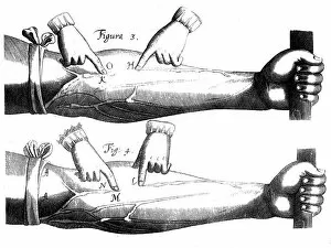
Background imageBlood Vessel Collection: Circulation of the blood, 1628

Circulation of the blood, 1628. English physician William Harvey (1578-1657) was the first to correctly describe the mechanism whereby blood is circulated in the body

Background imageBlood Vessel Collection: Illuminated Ships

Illuminated Ships
1957: Vessels of the Fleet are illuminated as seen from beneath the wing of a Ganet aircraft from HMS Ark Royal in Cromarty Firth

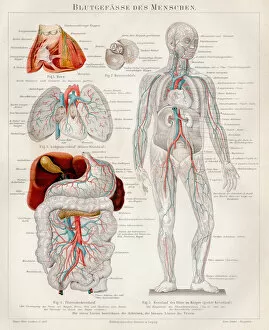
Background imageBlood Vessel Collection: Blood-vessels of the human engraving 1895

Blood-vessels of the human engraving 1895
Meyers Konversations-Lexikon. Ein Nachschlagewerk des allgemeinen Wissens, 5th edition 17 volumes Bibliographisches Institut - Leipzig 1895-1897

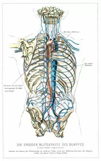
Background imageBlood Vessel Collection: Blood vessels anatomy engraving 1857

Blood vessels anatomy engraving 1857
Rank, johannes - The human being. 1 - 1894

Background imageBlood Vessel Collection: Nervous system anatomy engraving 1857

Nervous system anatomy engraving 1857
Rank, johannes - The human being. 1 - 1894

Background imageBlood Vessel Collection: Pulmonary alveolus

Pulmonary alveolus
Antique illustration of a Pulmonary alveolus

Background imageBlood Vessel Collection: Pulse oximetry

Pulse oximetry
Antique illustration of a Pulse oximetry

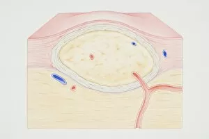
Background imageBlood Vessel Collection: Cross-section diagram of a non-cancerous tumour including a fibrous capsule

Cross-section diagram of a non-cancerous tumour including a fibrous capsule, tissue layer and blood vessel

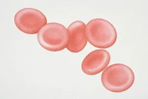
Background imageBlood Vessel Collection: Six red blood cells

Six red blood cells

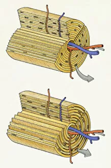
Background imageBlood Vessel Collection: Cross-section diagram of a human nerve fascicle, including a bundle of nerve fibres

Cross-section diagram of a human nerve fascicle, including a bundle of nerve fibres, blood vessels and myelin sheath

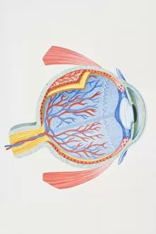
Background imageBlood Vessel Collection: Cross-section diagram of human eye

Cross-section diagram of human eye

Background imageBlood Vessel Collection: Cross-section diagram of tooth

Cross-section diagram of tooth

Background imageBlood Vessel Collection: Anchor and chain

Anchor and chain

Background imageBlood Vessel Collection: Two metal anchors

Two metal anchors

Background imageBlood Vessel Collection: Woman sat with leg outstretched to one side, other leg folded in, arm behind head, skeleton

Woman sat with leg outstretched to one side, other leg folded in, arm behind head, skeleton, lymphatic system illustration overlay

Background imageBlood Vessel Collection: Internal structure of human kidneys, with inset showing medulla

Internal structure of human kidneys, with inset showing medulla

Background imageBlood Vessel Collection: Breast showing rib, pectoral muscle, lung, fatty tissue, blood vessel, lobule, ampulla, nipple

Breast showing rib, pectoral muscle, lung, fatty tissue, blood vessel, lobule, ampulla, nipple, areola and milk duct, cross-section

Background imageBlood Vessel Collection: Illustration of formation of blood clot in series of four cross section illustrations showing

Illustration of formation of blood clot in series of four cross section illustrations showing blood oozing from blood vessel through wound before forming platelet plug

Background imageBlood Vessel Collection: Illustration of cross section of normal bone structure with blood vessels in central canal

Illustration of cross section of normal bone structure with blood vessels in central canal

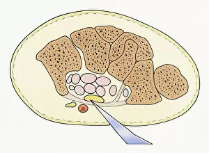
Background imageBlood Vessel Collection: Illustration showing cross section of severed median nerve in wrist caused by sharp piece of glass

Illustration showing cross section of severed median nerve in wrist caused by sharp piece of glass penetrating skin

Background imageBlood Vessel Collection: Digital illustration showing secondary tumour where cancerous cells have divided

Digital illustration showing secondary tumour where cancerous cells have divided and lodged in narrow blood vessel and surrounding tissue

Background imageBlood Vessel Collection: Digital illustration of blood vessel wall rupturing, primary tumour expanding

Digital illustration of blood vessel wall rupturing, primary tumour expanding, cells rupturing walls of blood vessels, enabling cancerous cells to detach and spread via blood flow

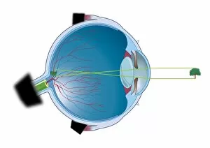
Background imageBlood Vessel Collection: Digital illustration of vertebrate eye using lens to focus light and form and image

Digital illustration of vertebrate eye using lens to focus light and form and image

Background imageBlood Vessel Collection: Digital illustration of invertebrate joint, left, and vertebrate joint, right

Digital illustration of invertebrate joint, left, and vertebrate joint, right

Background imageBlood Vessel Collection: Digital cross section illustration wound below human skin showing red and white blood cells and yell

Digital cross section illustration wound below human skin showing red and white blood cells and yell

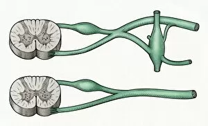
Background imageBlood Vessel Collection: Digital cross section illustration of sympathetic and parasympathetic nerves

Digital cross section illustration of sympathetic and parasympathetic nerves

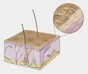
Background imageBlood Vessel Collection: Cross section illustration of structure of human skin

Cross section illustration of structure of human skin

Background imageBlood Vessel Collection: Illustration of low blood glucose response

Illustration of low blood glucose response

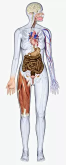
Background imageBlood Vessel Collection: Digital illustration of woman showing brain, oesophagus, heart, stomach, intestine, veins and muscle

Digital illustration of woman showing brain, oesophagus, heart, stomach, intestine, veins and muscle

Background imageBlood Vessel Collection: Digital illustration of young woman showing intestine, kidneys, heart, veins and muscle

Digital illustration of young woman showing intestine, kidneys, heart, veins and muscle

Background imageBlood Vessel Collection: Digital illustration showing arteries in human brain

Digital illustration showing arteries in human brain

Background imageBlood Vessel Collection: Digital illustration of human brain showing blood vessels, and areas of dead tissue highlighted in

Digital illustration of human brain showing blood vessels, and areas of dead tissue highlighted in red

Background imageBlood Vessel Collection: Digital illustration of autonomic nervous system responsible for automatic body functions

Digital illustration of autonomic nervous system responsible for automatic body functions

Background imageBlood Vessel Collection: Sequence of illustrations showing complete bone fracture and healing process

Sequence of illustrations showing complete bone fracture and healing process

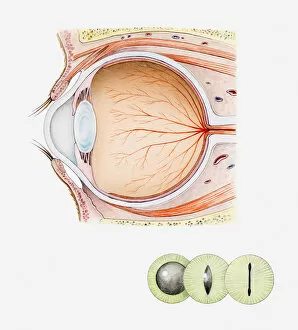
Background imageBlood Vessel Collection: Cross section iIllustration of eye of domestic cat with inset illustrations of green iris

Cross section iIllustration of eye of domestic cat with inset illustrations of green iris and black pupils

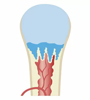
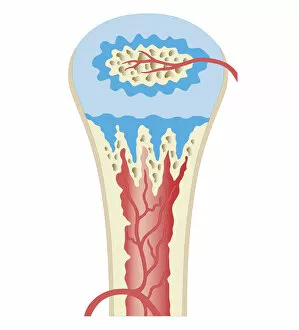
Background imageBlood Vessel Collection: Cross section biomedical illustration of long bone of newborn baby

Cross section biomedical illustration of long bone of newborn baby

Background imageBlood Vessel Collection: Cross section biomedical illustration of childhood development long bone

Cross section biomedical illustration of childhood development long bone

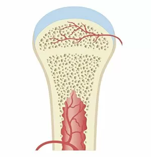
Background imageBlood Vessel Collection: Cross section biomedical illustration of fully developed long bone in adult

Cross section biomedical illustration of fully developed long bone in adult

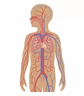
Background imageBlood Vessel Collection: Cross section biomedical illustration of cardiovascular system and respiratory system in child

Cross section biomedical illustration of cardiovascular system and respiratory system in child

Background imageBlood Vessel Collection: Cross section biomedical illustration of drug are released continuously from adhesive patch or gel

Cross section biomedical illustration of drug are released continuously from adhesive patch or gel on surface of skin surface passing through into blood vessel

Background imageBlood Vessel Collection: Cross section biomedical illustration of kidney transplant

Cross section biomedical illustration of kidney transplant


For sale as Licensed Images

Choose your image, Select your licence and Download the media

"Exploring the intricate network of blood vessels in our body: from brain to lungs, groin to face and scalp. " "Dive into the fascinating world of blood vessels with a 3D angiogram and a diagram showcasing their role in brain health. " "The vital connection between our lungs and bloodstream revealed through a detailed diagram highlighting the blood supply. " "Unveiling an artistic masterpiece from 1825, depicting male groin arteries - a testament to early anatomical studies. " "Discovering the superficial arteries and veins that grace our face and scalp, intricately illustrated for medical understanding. " "A mesmerizing artwork capturing the complexity of the blood coagulation cascade - an essential process for healing. " "Intriguing anatomy meets biology as we delve into colorful images showcasing various aspects function. " "Leonardo da Vinci's genius shines through pen and ink studies dating back to c1510, unraveling arm muscles alongside superficial vessels. " "With every beat, our human heart keeps us alive. Marvel at its beauty through captivating artwork that celebrates its strength. " "Journeying back in time with an exquisite 1825 artwork revealing head and chest arteries - a glimpse into historical medical knowledge. " "A close-up view of red blood cells captured by scanning electron microscopy showcases their unique structure within our bloodstream. " "Glimpsing at a healthy heart portrayed in stunning artwork; reminding us of this incredible organ's importance in maintaining life.