Brain Collection (#6)

"The intricate map of the brain: Exploring its motor and sensory pathways" Delving into the depths of our mind, we uncover the wonders of the brain's complex anatomy

Background imageBrain Collection: Brain Sketch

Brain Sketch
Beth Cai

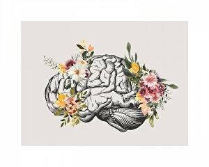
Background imageBrain Collection: Brain Bloom

Brain Bloom
Beth Cai

Background imageBrain Collection: You Are Not Your Thoughts No2

You Are Not Your Thoughts No2
Beth Cai

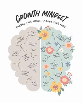
Background imageBrain Collection: Growth Mindset

Growth Mindset
Beth Cai

Background imageBrain Collection: Brain Bean

Brain Bean
Artem Pozdniakov

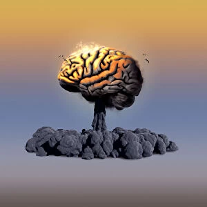
Background imageBrain Collection: Brain Explosion

Brain Explosion
Artem Pozdniakov

Background imageBrain Collection: Img 6858ps

Img 6858ps
Tove Höglund

Background imageBrain Collection: Img 6856ps

Img 6856ps
Tove Höglund

Background imageBrain Collection: Img 6890ps

Img 6890ps
Tove Höglund

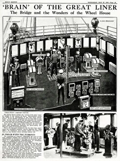
Background imageBrain Collection: Bridge and Wheel House of the Queen Mary

Bridge and Wheel House of the Queen Mary
The Bridge and Wheel House of the Queen Mary - Brain of the Great Liner Date: 1936

Background imageBrain Collection: Gentlemen In Conversation

Gentlemen In Conversation
A sketch comprising of a large group of top hat wearing gentlemen conversing in separate groups, relating to artists being the Brain of Paris Date: circa 1901

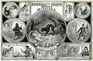
Background imageBrain Collection: Diseases of the Brain

Diseases of the Brain, Dreams, Delusions, Hallucinations and Nightmares Date: 1888

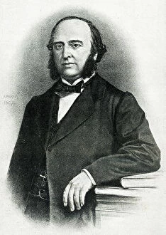
Background imageBrain Collection: Dr Paul Broca, French physician

Dr Paul Broca, French physician
Dr Paul Broca (1824-1880), French physician, anatomist and anthropologist, researcher on the area of the brain that deals with language. Date: circa 1850s

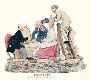
Background imageBrain Collection: Hochst Group

Hochst Group
An example of a Hochst group figurine, comprising of some men carefully tending to the appearance of the seated woman, one is holding a mirror

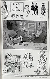
Background imageBrain Collection: Caricature illustrations of students, staff and academics at Cambridge University by H M Bateman

Caricature illustrations of students, staff and academics at Cambridge University by H M Bateman. Captioned, Cantabs in Caricature'

Background imageBrain Collection: Artistic Chaos

Artistic Chaos
Ivan Huang

Background imageBrain Collection: 20 Anniversary

20 Anniversary
DDiArte

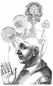
Background imageBrain Collection: The Cabalistic analysis of the mind and the senses, 1617-19 (engraving)

The Cabalistic analysis of the mind and the senses, 1617-19 (engraving)
539982 The Cabalistic analysis of the mind and the senses, 1617-19 (engraving) by English School, (17th century); (add.info.: The Cabalistic analysis of the mind and the senses)

Background imageBrain Collection: illustration from Traite de phrenologie humaine et comparee, 19th century (engraving)

illustration from Traite de phrenologie humaine et comparee, 19th century (engraving)
2625965 illustration from Traite de phrenologie humaine et comparee, 19th century (engraving) by French School, (19th century); (add.info)

Background imageBrain Collection: illustration from Traite de phrenologie humaine et comparee, 19th century (engraving)

illustration from Traite de phrenologie humaine et comparee, 19th century (engraving)
2625936 illustration from Traite de phrenologie humaine et comparee, 19th century (engraving) by French School, (19th century); (add.info)

Background imageBrain Collection: illustration from Traite de phrenologie humaine et comparee, 19th century (engraving)

illustration from Traite de phrenologie humaine et comparee, 19th century (engraving)
2625955 illustration from Traite de phrenologie humaine et comparee, 19th century (engraving) by French School, (19th century); (add.info)

Background imageBrain Collection: illustration from Traite de phrenologie humaine et comparee, 19th century (engraving)

illustration from Traite de phrenologie humaine et comparee, 19th century (engraving)
2625961 illustration from Traite de phrenologie humaine et comparee, 19th century (engraving) by French School, (19th century); (add.info)

Background imageBrain Collection: illustration from Traite de phrenologie humaine et comparee, 19th century (engraving)

illustration from Traite de phrenologie humaine et comparee, 19th century (engraving)
2625960 illustration from Traite de phrenologie humaine et comparee, 19th century (engraving) by French School, (19th century); (add.info)

Background imageBrain Collection: illustration from Traite de phrenologie humaine et comparee, 19th century (engraving)

illustration from Traite de phrenologie humaine et comparee, 19th century (engraving)
2625958 illustration from Traite de phrenologie humaine et comparee, 19th century (engraving) by French School, (19th century); (add.info)

Background imageBrain Collection: Medicine: 'The surgical operation'Painting by David Teniers II the young (1610-1690) (ec. flam)

Medicine: "The surgical operation"Painting by David Teniers II the young (1610-1690) (ec. flam. ) Sun
JLJ4685743 Medicine: " The surgical operation" Painting by David Teniers II the young (1610-1690) (ec.flam.) Sun

Background imageBrain Collection: Starvation, degrees of wasting (litho)

Starvation, degrees of wasting (litho)
5998374 Starvation, degrees of wasting (litho) by English School, (20th century); Private Collection; (add.info.: Starvation, degrees of wasting)

Background imageBrain Collection: The Extraction of the Stone of Madness (Cutting the Stone or The Cure of Folly) - Painting

The Extraction of the Stone of Madness (Cutting the Stone or The Cure of Folly) - Painting, 1475-1480
LRI4602862 The Extraction of the Stone of Madness (Cutting the Stone or The Cure of Folly) - Painting, 1475-1480 by Bosch, Hieronymus (c.1450-1516); Prado, Madrid

Background imageBrain Collection: Anatomy of a horse. 1792 (engraving)

Anatomy of a horse. 1792 (engraving)
7235905 Anatomy of a horse. 1792 (engraving) by Dodd, Daniel (fl.1760-90) (after); (add.info.: Anatomy of a horse. The head, brains, stomach, pancreas, and generative parts)

Background imageBrain Collection: DDE-90038223

DDE-90038223
Grooved Brain Coral (Diploria labyrinthiformis) Coral Reef Island, Belize Barrier Reef. Second largest barrier reef system in the world. Belize, Central America Date: 25/07/2011

Background imageBrain Collection: DDE-90035053

DDE-90035053
Russia, Moscow Oblast, Moscow, Sparrow Hills-area, elevated city view with the Russian Academy of Sciences, RAN, building, dusk Date: 05/01/2008

Background imageBrain Collection: DDE-90027106

DDE-90027106
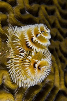
Christmas Tree Worm (Spirobranchus giganteus) on Grooved Brain Coral (Diploria labyrinthiformis), Bonaire, Netherlands Antilles, Caribbean Date: 08/11/2011

Background imageBrain Collection: DDE-90027087

DDE-90027087
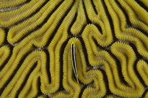
Grooved Brain Coral (Diploria labyrinthiformis) & Yellownose Goby (Elacatinus randalii), Bonaire, Netherlands Antilles, Caribbean Date: 07/11/2007

Background imageBrain Collection: DDE-90027032

DDE-90027032
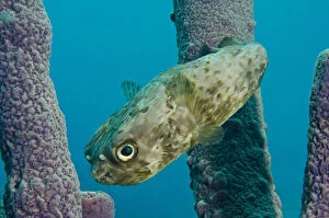
Balloonfish (Diodon holocanthus) in Stove-pipe Sponge (Aplysina archeri), Bonaire, Netherlands Antilles, Caribbean Date: 07/11/2007

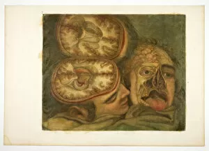
Background imageBrain Collection: Cranial Dissection, plate five from Anatomy of the Head, in Printed Paintings, 1748

Cranial Dissection, plate five from Anatomy of the Head, in Printed Paintings, 1748

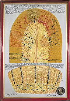
Background imageBrain Collection: Histological shema of the cerebellum (cerebellum) and its structure

Histological shema of the cerebellum (cerebellum) and its structure. Original drawing by Santiago Ramon y Cajal
AIS5376434 Histological shema of the cerebellum (cerebellum) and its structure. Original drawing by Santiago Ramon y Cajal (1852-1934), histologist and neuroscientist

Background imageBrain Collection: Surgery: A doctor discarding a patients cranial box. Miniature in 'Liber notabilium Philippi

Surgery: A doctor discarding a patients cranial box. Miniature in "Liber notabilium Philippi septimi francorum
JLJ4673994 Surgery: A doctor discarding a patients cranial box. Miniature in " Liber notabilium Philippi septimi francorum regis, a libris Galieni extractus" written by Gui (Guy) of Pavia

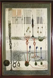
Background imageBrain Collection: Diagram of the structure of the mammalian retina, original drawing by Santiago Ramon y Cajal

Diagram of the structure of the mammalian retina, original drawing by Santiago Ramon y Cajal (1852-1934) (drawing)
AIS5384914 Diagram of the structure of the mammalian retina, original drawing by Santiago Ramon y Cajal (1852-1934) (drawing) by Ramon y Cajal, Santiago (1852-1934); © Iberfoto

Background imageBrain Collection: Anatomy and surgical boards: crane box showing the brains of two men barks

Anatomy and surgical boards: crane box showing the brains of two men barks
JLJ4619521 Anatomy and surgical boards: crane box showing the brains of two men barks. In " Liber notabilium Philippi septimi francorum regis

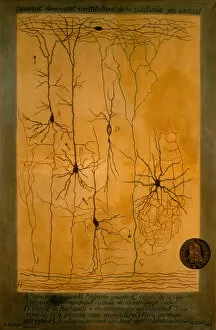
Background imageBrain Collection: Histological diagram of the constituent elements of the cerebral cortex (grey matter of the brain)

Histological diagram of the constituent elements of the cerebral cortex (grey matter of the brain)
AIS5385499 Histological diagram of the constituent elements of the cerebral cortex (grey matter of the brain), the neurons are represented, 19th-20th century (drawing) by Ramon y Cajal

Background imageBrain Collection: Anatomy and surgical boards: crane box (mans brain). In 'Liber notabilium Philippi septimi

Anatomy and surgical boards: crane box (mans brain). In "Liber notabilium Philippi septimi francorum regis
JLJ4619554 Anatomy and surgical boards: crane box (mans brain). In " Liber notabilium Philippi septimi francorum regis, a libris Galieni extratus" by Gui de Pavia circa 1345

Background imageBrain Collection: Diplodocus (colour litho)

Diplodocus (colour litho)
5995626 Diplodocus (colour litho) by Stokoe, W. J.(fl.1910-40); Private Collection; (add.info.: Diplodocus. 80 feet long; very weak laterally, with a brain no bigger than a hens egg)

Background imageBrain Collection: Brain Ashton Celebrates

Brain Ashton Celebrates France V England, Rugby World CupFrance 07 France V England, World Cup Semi-Final Stade France, Paris, France 13 October 2007 Date: 13 October 2007

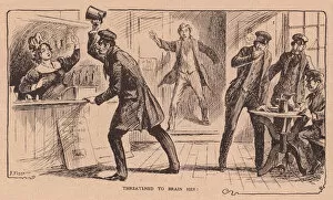
Background imageBrain Collection: Threatened to brain her! (litho)

Threatened to brain her! (litho)
7152411 Threatened to brain her! (litho) by English School, (20th century); Private Collection; (add.info.: Threatened to brain her)

Background imageBrain Collection: Section of Skull and Brain (litho)

Section of Skull and Brain (litho)
7180480 Section of Skull and Brain (litho) by English School, (20th century); Private Collection; (add.info.: Section of Skull and Brain)

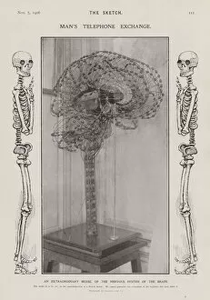
Background imageBrain Collection: Model of the nervous system of the human brain (b / w photo)

Model of the nervous system of the human brain (b / w photo)
7185863 Model of the nervous system of the human brain (b/w photo) by English School, (20th century); Private Collection; (add.info.: Model of the nervous system of the human brain)

Background imageBrain Collection: Saggital section of the human brain. 19th century (engraving)

Saggital section of the human brain. 19th century (engraving)
7212166 Saggital section of the human brain. 19th century (engraving) by Jacob, Nicolas Henri (1782-1871); (add.info.: Saggital section of the human brain)

Background imageBrain Collection: Model of the conjoined brains of Ladan and Laleh Bijani used by Dr. Ben Carson, 2003

Model of the conjoined brains of Ladan and Laleh Bijani used by Dr. Ben Carson, 2003
African-American neurosurgeon, author, and politician Benjamin Solomon Carson Sr. (born 1951) served as the 17th United States Secretary of Housing and Urban Development from 2017 to 2021

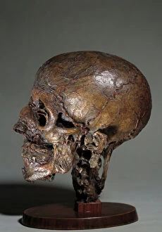
Background imageBrain Collection: Human anatomical head made by means of wax injections. 18th century Private collection

Human anatomical head made by means of wax injections. 18th century Private collection
JLJ4586758 Human anatomical head made by means of wax injections. 18th century Private collection by Unknown artist, (18th century); Private Collection; (add.info)


For sale as Licensed Images

Choose your image, Select your licence and Download the media

"The intricate map of the brain: Exploring its motor and sensory pathways" Delving into the depths of our mind, we uncover the wonders of the brain's complex anatomy. The Motor homunculus model reveals a distorted representation of our body, with certain regions magnified to depict their significance in movement control. Similarly, the Sensory homunculus showcases how different areas are dedicated to processing various sensations, emphasizing their importance in perceiving touch, pain, and temperature. Transporting us back in time, an engraving from 1895 unveils a vintage depiction anatomy. This historical artwork reminds us of the continuous quest for understanding this enigmatic organ that has captivated scientists for centuries. Zooming closer under a microscope lens, we witness the delicate beauty within cerebellum tissue through a light micrograph. Its intricate structure plays a crucial role in coordinating movements and maintaining balance. The Brain fibres captured by DTI MRI scan C017 / 7099 provide insight into its connectivity network. These fibers act as highways transmitting information between different regions - like bridges connecting distant lands. An inferior view offers another perspective on human brain anatomy. It unravels hidden complexities beneath our surface thoughts – showcasing structures responsible for memory formation, language processing, and emotional regulation. Artistic imagination breathes life into white matter fibres and brains through artwork C015 / 1930. This captivating portrayal merges science with creativity to portray both scientific accuracy and aesthetic appeal simultaneously. Venturing deeper within this mysterious realm is an exploration blood vessels using a stunning 3D angiogram C007 /1981. These intricate networks supply oxygen-rich blood to nourish every nook and cranny of this vital organ. Returning once again to explore motor and sensory functions intricately mapped out by homunculi models – these visual representations highlight how specific regions control precise movements or perceive distinct sensations throughout our body A masterpiece artwork captures Medulla oblongata in the brain, showcasing its vital role in controlling essential functions