Brain Collection (#9)

"The intricate map of the brain: Exploring its motor and sensory pathways" Delving into the depths of our mind, we uncover the wonders of the brain's complex anatomy

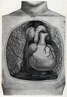
Background imageBrain Collection: aneurysm rupture (engraving) 1888

aneurysm rupture (engraving) 1888
ELD5618905 aneurysm rupture (engraving) 1888 by French School, (19th century); Private Collection; (add.info.: Engraving from " The Illustrated Medicine" ); Photo eIsadora; French

Background imageBrain Collection: Human sensory system connects to the brain - in 'L

Human sensory system connects to the brain - in "L
LSE4095867 Human sensory system connects to the brain - in " L homme de Rene Descartes et la formation du fetus", Paris, 1677 by Unknown Artist

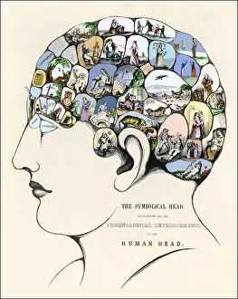
Background imageBrain Collection: Phrenology: Phrenological development. 19th century engraving

Phrenology: Phrenological development. 19th century engraving
XEE4410539 Phrenology: Phrenological development. 19th century engraving. by Unknown Artist, (19th century); Private Collection; (add.info.: Phrenology: Phrenological development)

Background imageBrain Collection: Surgical board representing different types of surgical instruments, hammer, scallop

Surgical board representing different types of surgical instruments, hammer, scallop
GIA4793009 Surgical board representing different types of surgical instruments, hammer, scallop, knives, for the removal of the brain

Background imageBrain Collection: Skull cut. Anatomical board of 'De dissectione partium corporis humani lbri

Skull cut. Anatomical board of "De dissectione partium corporis humani lbri
GIA4772236 Skull cut. Anatomical board of " De dissectione partium corporis humani lbri tres..." Carlo Stefano Riverio, Paris 1545 by Unknown Artist

Background imageBrain Collection: Cover of 'Le Lire', number 278, Satirique en Colours

Cover of "Le Lire", number 278, Satirique en Colours
ICA4873234 Cover of " Le Lire", number 278, Satirique en Colours, 1900_3_3: L operation du trepan - Medical, Trepanation - Medicine - Illustration by Abel Faivre (1867-1945) by Faivre

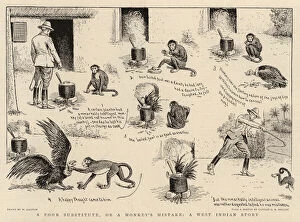
Background imageBrain Collection: A Poor Substitute, or a Monkeys Mistake, a West Indian Story (litho)

A Poor Substitute, or a Monkeys Mistake, a West Indian Story (litho)
1039434 A Poor Substitute, or a Monkeys Mistake, a West Indian Story (litho) by Ralston, William (1848-1911) (after); Private Collection; (add.info.: A Poor Substitute, or a Monkeys Mistake)

Background imageBrain Collection: Can I Play with Madness

Can I Play with Madness
Petri Damstén

Background imageBrain Collection: Every Last Drop of Ideas

Every Last Drop of Ideas
Petri Damstén

Background imageBrain Collection: Egghead

Egghead
Petri Damstén

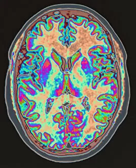
Background imageBrain Collection: MRI scan of brain

MRI scan of brain
body part, brain, bright, colour, conditions, medical, medical scan, medical treatment, mri scan, multi coloured, one person, someone, technology, vertical, photography, brain artwork, 56961661

Background imageBrain Collection: Model of human brain, studio shot

Model of human brain, studio shot
intelligence, anatomical model, anatomy, black background, brain, complexity, healthcare and medicine, horizontal, no people, pattern, single object, studio shot, wisdom, brain artwork, 114850733

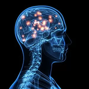
Background imageBrain Collection: Brain activity, artwork

Brain activity, artwork
Brain activity, computer artwork

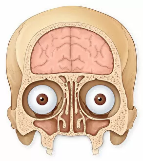
Background imageBrain Collection: Normal coronal section of the skull and brain showing the coronal sinuses

Normal coronal section of the skull and brain showing the coronal sinuses
Brain, Anatomy, Diagram, Illustration, Bone, Skull, Medical, Eyeball, Biology, See Through, Cut Out, White Background, Eye, Vertical, Artwork, Color Image, Front View, Human Skull

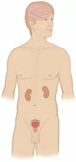
Background imageBrain Collection: Normal front view of a man showing the endocrine system including the brain

Normal front view of a man showing the endocrine system including the brain
Brain, Anatomy, Diagram, Illustration, Naked, Medical, Biology, See Through, Cut Out, White Background, Vertical, Artwork, Male Likeness, Color Image, Front View, Healthcare And Medicine

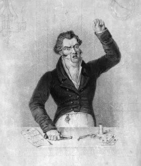
Background imageBrain Collection: THE GAMBLER C1810

THE GAMBLER C1810
He loses all... till madness seizes on his fevrish brain A gentleman gambler, deep in debt, raises his fist in impotent rage. Date: circa 1810

Background imageBrain Collection: Anatomy - Spine and Brain

Anatomy - Spine and Brain
Detailed diagram of the brain and spine Date: 1768

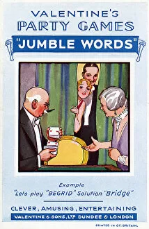
Background imageBrain Collection: GAME / JUMBLE WORDS 1930S

GAME / JUMBLE WORDS 1930S
A delightful game for young and old, taxing the brain by asking you to guess Come down smiling to BARK FEAST and other anagrammatic enigmas Date: 1930s

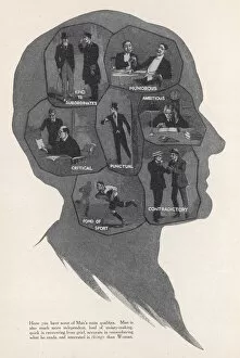
Background imageBrain Collection: WOMENS RIGHTS / MIND

WOMENS RIGHTS / MIND
The main qualities of a Man 1 of 2 Date: 1913

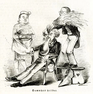
Background imageBrain Collection: SURGERY CURES STUPIDITY

SURGERY CURES STUPIDITY
SATIRE:RESULTS OF ETHER - surgery as a cure-all: young mans stupidity cured by replacing straw head- stuffing with a brain! Date: 1847

Background imageBrain Collection: Valentines Day, mid 19th century. Creator: J Brain

Valentines Day, mid 19th century. Creator: J Brain
Valentines Day, mid 19th century. A young man is revealed behind a curtain, reading a letter, while his sisters giggle and point

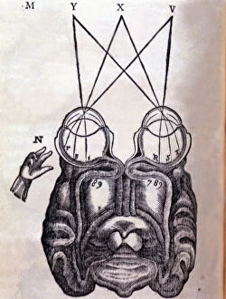
Background imageBrain Collection: Schematic drawing on the functioning of the brain and human ocular system, engraving

Schematic drawing on the functioning of the brain and human ocular system, engraving published in the book Principia Philosophiae, 1677

Background imageBrain Collection: Prague, c1850. Artist: Albert Henry Payne

Prague, c1850. Artist: Albert Henry Payne
Prague, c1850. From Paynes Universum, or, Pictorial world : being a collection of engravings of views in all countries, portraits of great men, and specimens of works of art

Background imageBrain Collection: Wassili Blagennoi or the Cathedral of St. Basil Moscow, c1850. Artist: Albert Henry Payne

Wassili Blagennoi or the Cathedral of St. Basil Moscow, c1850. Artist: Albert Henry Payne
Wassili Blagennoi or the Cathedral of St. Basil Moscow, c1850. After D. Bydgoszcz. From Paynes Universum, or, Pictorial world : being a collection of engravings of views in all countries

Background imageBrain Collection: Sir Benjamin Collins Brodie, 19th century. Artist: J Brain

Sir Benjamin Collins Brodie, 19th century. Artist: J Brain
Sir Benjamin Collins Brodie (1783-1862), English physiologist and surgeon, 19th century. Brodie pioneered research into bone and joint disease

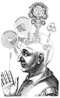
Background imageBrain Collection: The Cabalistic analysis of the mind and the senses, 1617

The Cabalistic analysis of the mind and the senses, 1617. This attributed different functions to different regions of the brain. From Ultriusque cosmi...historia by Robert Fludd. (Oppenheim, 1617-1619
Background imageBrain Collection: Franz Joseph Gall, German physician and founder of Phrenology, c1790

Franz Joseph Gall, German physician and founder of Phrenology, c1790
Franz Joseph Gall, German physician and founder of Phrenology, c1820. Phrenology was a theory, propounded by Gall (1758-1828) in around 1800

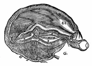
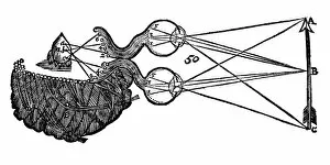
Background imageBrain Collection: Rene Descartes diagram of the human brain and eye, 1692

Rene Descartes diagram of the human brain and eye, 1692. From Opera Philosophica by Rene Descartes. (Frankfurt-am-Main, 1692). Originally published in his Tractatus de homine. (Paris, 1664)

Background imageBrain Collection: Rene Descartes idea of vision, showing the function of the eye, optic nerve and brain, 1692

Rene Descartes idea of vision, showing the function of the eye, optic nerve and brain, 1692. From Opera Philosophica by Rene Descartes. (Frankfurt-am-Main, 1692)

Background imageBrain Collection: Descartes idea of vision, 1692

Descartes idea of vision, 1692
Descartes (1596-1650) idea of vision, [1692]. The passage of nervous impulses from the eye to the pineal gland and so to the muscles. From Rene Descartes Opera Philosophica (Tractatus de homine), 1692

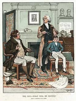
Background imageBrain Collection: Phrenology, 1820 (1886). Artist: Frank Dadd

Phrenology, 1820 (1886). Artist: Frank Dadd
Phrenology, 1820 (1886). Measuring the bumps on a boys head to assess his future. On the wall in the background is a portrait of of Franz Josef Gall (1757-1828)

Background imageBrain Collection: Phrenology chart, showing presumed areas of activity of the brain, c1920

Phrenology chart, showing presumed areas of activity of the brain, c1920. Phrenology was a theory, propounded by the German physician Franz Joseph Gall in around 1800

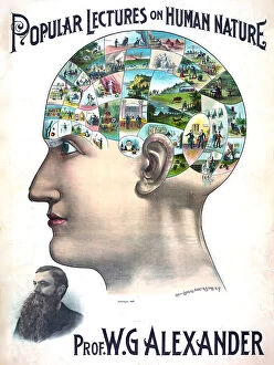
Background imageBrain Collection: Popular lectures on human nature by Professor W. G. Alexander poster of man with ideas in his

Popular lectures on human nature by Professor W. G. Alexander poster of man with ideas in his brain ca. 1895
Popular lectures on human nature by Professor W.G. Alexander poster of man with ideas in his brain ca. 1895

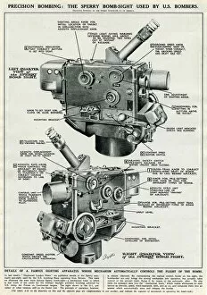
Background imageBrain Collection: Sperry bomb-sight apparatus 1944

Sperry bomb-sight apparatus 1944
Diagram of a Sperry bomb-sight O-1 used by the United States Army Air Forces (USaF) bombers in World War Two. By manipulating the various control knobs

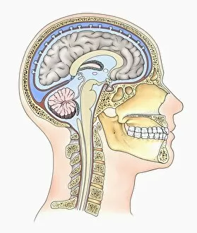
Background imageBrain Collection: Lateral cross section through the skull showing the brain with intermediate layers (meninges)

Lateral cross section through the skull showing the brain with intermediate layers (meninges) highlighted

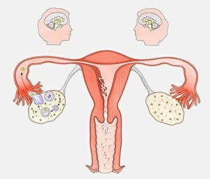
Background imageBrain Collection: Diagram showing the interaction between female sexual organs and the brain, on one side

Diagram showing the interaction between female sexual organs and the brain, on one side, the normal reproductive cycle, and on the other, the effect of the contraceptive pill

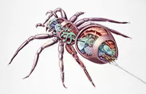
Background imageBrain Collection: Spider (Araneae), internal anatomy, cross-section

Spider (Araneae), internal anatomy, cross-section

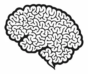
Background imageBrain Collection: Digital cartoon of human brain showing cerebrum

Digital cartoon of human brain showing cerebrum

Background imageBrain Collection: Digital illustration of showing front view of human brain

Digital illustration of showing front view of human brain

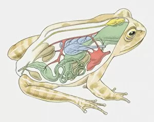
Background imageBrain Collection: Illustration of anatomy of frog

Illustration of anatomy of frog

Background imageBrain Collection: Illustration of female human body showing skeleton, brain, heart, lungs, digestive system

Illustration of female human body showing skeleton, brain, heart, lungs, digestive system, and uterus

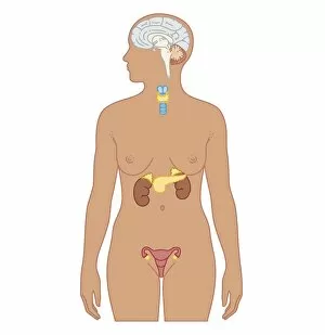
Background imageBrain Collection: Cross section biomedical illustration of endocrine system in adult female

Cross section biomedical illustration of endocrine system in adult female

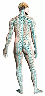
Background imageBrain Collection: Diagram of human nervous system, posterior view

Diagram of human nervous system, posterior view

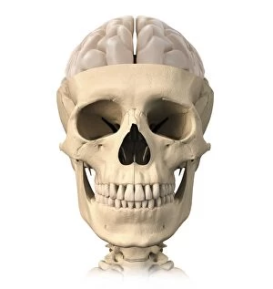
Background imageBrain Collection: Anatomy of human skull, cutaway view with half brain showing

Anatomy of human skull, cutaway view with half brain showing, front view

Background imageBrain Collection: Human brain anatomy, superior view

Human brain anatomy, superior view

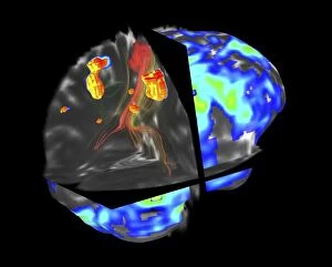
Background imageBrain Collection: Brain tumour, fMRI and tractography C017 / 7102

Brain tumour, fMRI and tractography C017 / 7102
Brain tumour, fMRI and tractography. Combined functional magnetic resonance imaging (fMRI, blue and green) and tractography (yellow and red) imaging of a brain with a tumour (upper left)

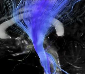
Background imageBrain Collection: Corticospinal tract, DTI MRI scan C017 / 7046

Corticospinal tract, DTI MRI scan C017 / 7046
Corticospinal tract. Sagittal 3D diffusion tensor imaging (DTI) magnetic resonance imaging (MRI) scan of nerve pathways (blue) in the brains corticospinal tract

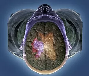
Background imageBrain Collection: Stroke, MRI and 3D CT scans C016 / 6419

Stroke, MRI and 3D CT scans C016 / 6419
Stroke. Coloured composite image of a magnetic resonance imaging (MRI) scan of the brain and 2D and 3D computed tomography (CT)


For sale as Licensed Images

Choose your image, Select your licence and Download the media

"The intricate map of the brain: Exploring its motor and sensory pathways" Delving into the depths of our mind, we uncover the wonders of the brain's complex anatomy. The Motor homunculus model reveals a distorted representation of our body, with certain regions magnified to depict their significance in movement control. Similarly, the Sensory homunculus showcases how different areas are dedicated to processing various sensations, emphasizing their importance in perceiving touch, pain, and temperature. Transporting us back in time, an engraving from 1895 unveils a vintage depiction anatomy. This historical artwork reminds us of the continuous quest for understanding this enigmatic organ that has captivated scientists for centuries. Zooming closer under a microscope lens, we witness the delicate beauty within cerebellum tissue through a light micrograph. Its intricate structure plays a crucial role in coordinating movements and maintaining balance. The Brain fibres captured by DTI MRI scan C017 / 7099 provide insight into its connectivity network. These fibers act as highways transmitting information between different regions - like bridges connecting distant lands. An inferior view offers another perspective on human brain anatomy. It unravels hidden complexities beneath our surface thoughts – showcasing structures responsible for memory formation, language processing, and emotional regulation. Artistic imagination breathes life into white matter fibres and brains through artwork C015 / 1930. This captivating portrayal merges science with creativity to portray both scientific accuracy and aesthetic appeal simultaneously. Venturing deeper within this mysterious realm is an exploration blood vessels using a stunning 3D angiogram C007 /1981. These intricate networks supply oxygen-rich blood to nourish every nook and cranny of this vital organ. Returning once again to explore motor and sensory functions intricately mapped out by homunculi models – these visual representations highlight how specific regions control precise movements or perceive distinct sensations throughout our body A masterpiece artwork captures Medulla oblongata in the brain, showcasing its vital role in controlling essential functions