
Budding Collection (#4)
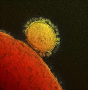
"Budding: A Symphony of Life's Creative Flourish" In the quiet corners of nature, a budding yeast cell delicately emerges, silently practicing its own harmonious growth


For sale as Licensed Images
Choose your image, Select your licence and Download the media
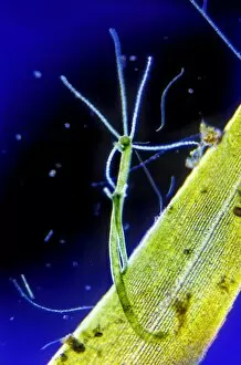
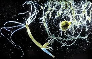
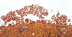
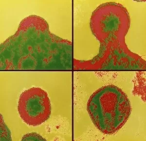
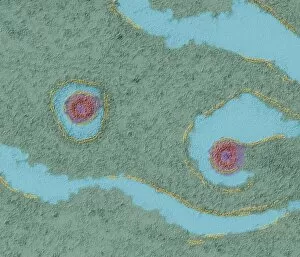
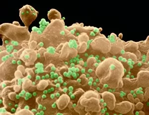
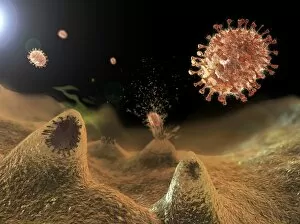
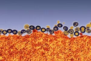
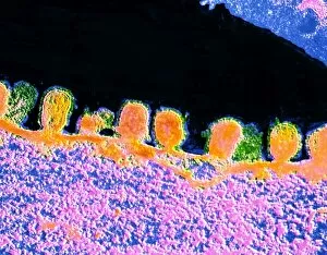
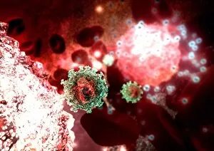
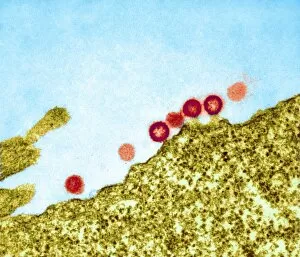
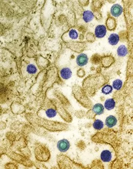
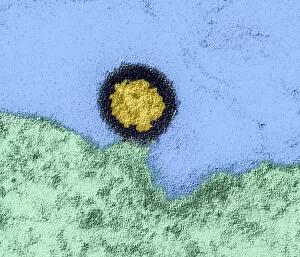
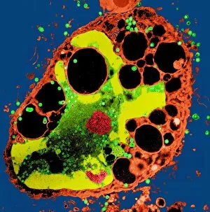
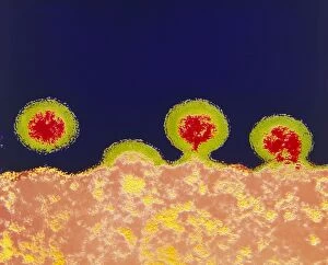
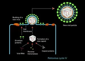
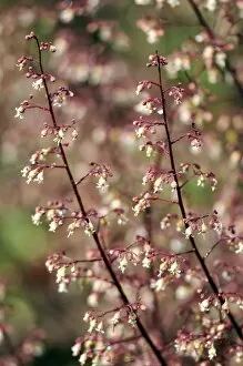
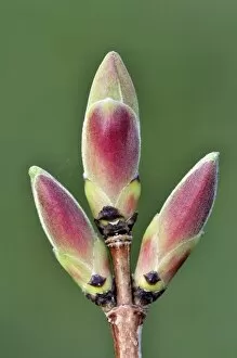
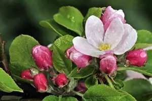
"Budding: A Symphony of Life's Creative Flourish" In the quiet corners of nature, a budding yeast cell delicately emerges, silently practicing its own harmonious growth. Like a musician perfecting their craft, it embraces the art with grace and precision. Meanwhile, Candida fungus reveals its intricate beauty under the watchful eye of a scanning electron microscope. Its delicate structures captivate our imagination as we marvel at the wonders hidden within this tiny organism. The world extends beyond microorganisms; even viruses partake in this captivating process. Rift Valley fever virus and vesicular stomatitis virus are captured through transmission electron microscopy, showcasing their unique way of propagating life. Herpes simplex viruses and AIDS viruses also engage in an enchanting dance as they bud from infected cells. These microscopic spectacles remind us that life persists even in the most unexpected places. Amidst these fascinating glimpses into nature's creativity, Candida albicans yeast takes center stage once again. Scanning electron microscopy unveils its textured surface like an artist's canvas waiting to be explored. Beyond the realm of biology lies another form – cultivated roses blooming with vibrant colors and intoxicating fragrances. J. C Vickery Advertisement beckons us to indulge in these floral masterpieces that symbolize love and beauty. And finally, Samuelson's lawn mowing machines invite us to witness yet another type – one where landscapes transform under skilled hands into meticulously manicured works of art. From microscopic organisms to grand gardens, "budding" encapsulates life's continuous cycle of creation and renewal. It reminds us that every living thing has within itself the potential for growth and transformation – a testament to nature's boundless ingenuity.