Cell Collection (#94)

"Exploring the Intricacies of Life: Unveiling the Wonders of Cells" Delving into the microscopic world

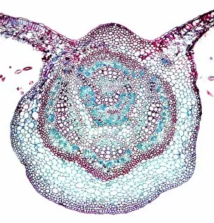
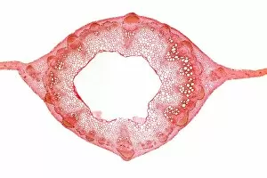
Background imageCell Collection: Sycamore leaf vein, light micrograph

Sycamore leaf vein, light micrograph
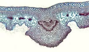
Sycamore leaf vein. Light micrograph of a section through the midrib (vein) of a leaf from a sycamore (Acer pseudoplatanus) tree

Background imageCell Collection: Bean pod, light micrograph

Bean pod, light micrograph
Bean pod. Light micrograph of a section through the seed pod of a bean (Phaseolus sp.) plant. The pericarp (outer covering)

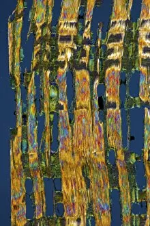
Background imageCell Collection: African teak woody stem, light micrograph

African teak woody stem, light micrograph
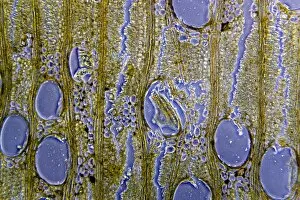
African teak woody stem. Polarised light micrograph of a section through the stem of an African teak (Pericopsis elata) plant, showing its diffuse-porous xylem tissue

Background imageCell Collection: Venus flytrap leaf, light micrograph

Venus flytrap leaf, light micrograph
Venus flytrap leaf. Light micrograph of a section through the leaf of a Venus flytrap (Dionaea sp.), showing the digestive glands that line its interior

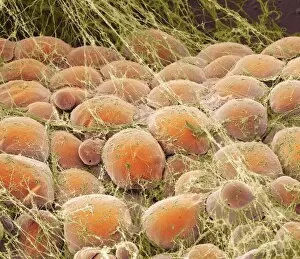
Background imageCell Collection: Fat tissue, SEM

Fat tissue, SEM
Fat tissue. Coloured scanning electron micrograph (SEM) of fat cells (adipocytes, round) surrounded by fine strands of supportive connective tissue

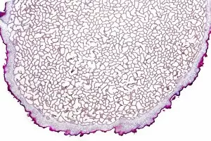
Background imageCell Collection: Earth ball fungus, light micrograph

Earth ball fungus, light micrograph
Earth ball fungus. Light micrograph of a section through tissue from the fruiting body of an earth ball (Scleroderma vulgare) mushroom

Background imageCell Collection: Plum pocket infection, light micrograph

Plum pocket infection, light micrograph
Plum pocket infection. Light micrograph of a section through a plum gall caused by the plum pocket fungus (Taphrina pruni)
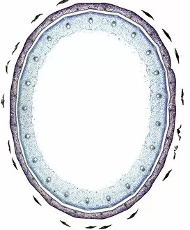
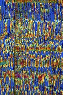
Background imageCell Collection: Beech tree stem, light micrograph

Beech tree stem, light micrograph
Beech tree stem. Polarised light micrograph of a section through the woody stem of a common beech tree (Fagus sylvatica), showing diffuse-porous xylem vessels (dark blue)

Background imageCell Collection: Female pine cone scales, light micrograph

Female pine cone scales, light micrograph
Female pine cone scales. Light micrograph of a section through two scales from a young ovulate female Scots pine (Pinus sylvestris) cone, bearing ovules (large, oval)

Background imageCell Collection: Geranium stem, light micrograph

Geranium stem, light micrograph
Geranium stem. Light micrograph of a section through a young stem of a geranium (Pelargonium sp.) plant. The outer stem is covered with a thin epidermis (red) which has stomata

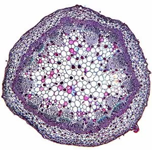
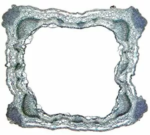
Background imageCell Collection: Sweet pea stem, light micrograph

Sweet pea stem, light micrograph
Sweet pea stem. Light micrograph of a section through the hollow stem of a sweet pea (Lathyrus odoratus) plant, showing a ring of vascular bundles

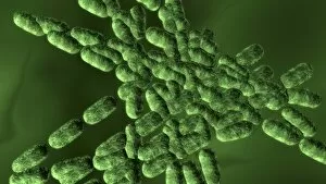
Background imageCell Collection: Dividing bacteria, computer artwork

Dividing bacteria, computer artwork
Bacteria dividing, computer artwork

Background imageCell Collection: Rust fungus in a leaf, light micrograph

Rust fungus in a leaf, light micrograph
Rust fungus in a leaf. Light micrograph of a section through a wheat leaf infected with the parasitic Puccinia graminis rust

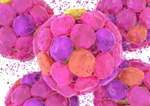
Background imageCell Collection: Islets of Langerhans cells, artwork

Islets of Langerhans cells, artwork
Islets of Langerhans cells, computer artwork. These cells (large, pink) of the pancreas release hormones (dots) into the blood

Background imageCell Collection: Shepherds purse fruit, light micrograph

Shepherds purse fruit, light micrograph
Shepherds purse fruit. Light micrograph of a section through a seed pod from a shepherds purse (Capsella bursa-pastoris) plant. The remains of the stigma can be seen (top centre)

Background imageCell Collection: Sage stem, light micrograph

Sage stem, light micrograph
Sage stem. Light micrograph of a section through a primary stem of a scarlet sage (Salivia splendens) plant. The outer stem is covered with a thin epidermis (green) that contains stomata

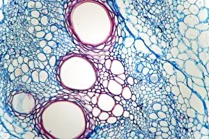
Background imageCell Collection: Rhubarb root, light micrograph

Rhubarb root, light micrograph
Rhubarb root. Light micrograph of a section through the root of a rhubarb (Rheum sp.) plant. The primary cortex has been shed by the formation of a circular meristem, the periderm

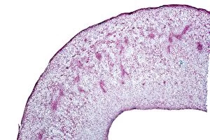
Background imageCell Collection: Sycamore leaf in autumn, micrograph

Sycamore leaf in autumn, micrograph
Sycamore leaf in autumn. Light micrograph of a section through the stem of a leaf from a sycamore (Acer pseudoplantanus) tree in autumn

Background imageCell Collection: Lime tree stem wound, light micrograph

Lime tree stem wound, light micrograph
Lime tree stem wound. Light micrograph of a section through the wounded stem of a lime tree (Tilia europaea). The outer epidermis has been shed and replaced by a layer of cork (dark red)

Background imageCell Collection: Penicillium mould, light micrograph

Penicillium mould, light micrograph
Penicillium mould. Light micrograph of a section through an orange rind infected with Penicillium sp. blue mould fungus, showing fungal hyphae and conidiophores (pink and purple) with spores (red)

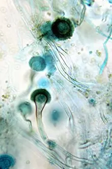
Background imageCell Collection: Brown mould fungus, light micrograph

Brown mould fungus, light micrograph
Brown mould fungus. Light micrograph of a section through decaying plant tissue infected with brown mould fungus (Aspergillus glaucum)

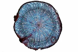
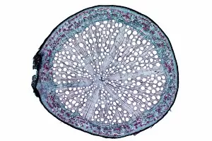
Background imageCell Collection: Oak root, light micrograph

Oak root, light micrograph
Oak root. Light micrograph of a section through a secondary root from an oak (Quercus sp.) tree. The primary cortex (outer layer) has been shed by the formation of a circular meristem, the periderm

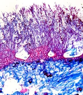
Background imageCell Collection: Boletus fungus, light micrograph

Boletus fungus, light micrograph
Boletus fungus. Light micrograph of a section through tissue from the fruiting body of a Boletus sp. mushroom, showing the hymenium (dark red) of the fruiting body

Background imageCell Collection: Downy mildew infection, light micrograph

Downy mildew infection, light micrograph
Downy mildew infection. Light micrograph of a section through a grapevine (Vitis sp.) leaf infected with downy mildew (Plasmopara viticola)

Background imageCell Collection: African mahogany stem, light micrograph

African mahogany stem, light micrograph
African mahogany stem. Polarised light micrograph of a section through the stem of an African mahogany (Khaya sp.) tree. The multicoloured cells are fibres, and fibre tracheids, which carry water

Background imageCell Collection: White bryony stem, light micrograph

White bryony stem, light micrograph
White bryony stem. Light micrograph of a transverse section through the stem of a white bryony (Bryonia alba) plant, showing a single collateral vascular bundle

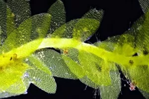
Background imageCell Collection: Mnium moss, light micrograph

Mnium moss, light micrograph
Mnium moss. Light micrograph of Mnium punctatum moss showing its leaves, which are composed of a single layer of cells full of chloroplasts

Background imageCell Collection: Beech tree leaves, light micrograph

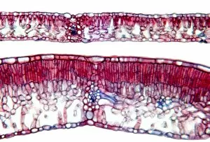
Beech tree leaves, light micrograph
Beech tree leaves. Light micrograph of a section through two leaves from different parts of a common beech tree (Fagus sylvatica)

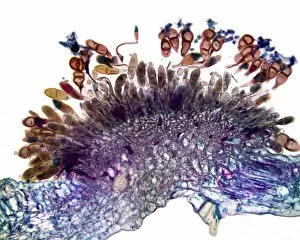
Background imageCell Collection: Hollyhock rust fungus in a leaf

Hollyhock rust fungus in a leaf. Light micrograph of a section through a hollyhock (Alcea sp.) leaf infected with the parasitic Puccinia malvacearum rust, showing the teliospores (spores)

Background imageCell Collection: Orange fruit, light micrograph

Orange fruit, light micrograph
Orange fruit. Light micrograph of a section through the rind of a fruit from a bitter orange (Citrus aurantium) tree, showing one of the many spherical oil-secreting glands

Background imageCell Collection: Mnium moss

Mnium moss. Close-up of Mnium punctatum moss showing its leaves, which are composed of a single layer of cells full of chloroplasts

Background imageCell Collection: Female pine cone, light micrograph

Female pine cone, light micrograph
Female pine cone. Light micrograph of a section through a young ovulate female cone from a Scots pine (Pinus sylvestris) tree, showing ovules (oval, light pink)

Background imageCell Collection: Shining hookeria moss, light micrograph

Shining hookeria moss, light micrograph
Shining Hookeria moss. Dark field light micrograph of a section through shining hookeria (Hookeria luscens) moss, showing the stem and leaves

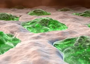
Background imageCell Collection: Osteoclast bone cells, artwork

Osteoclast bone cells, artwork
Osteoclast bone cells. Computer artwork of normal osteoclasts (green) in the lacunae (spaces) of bone tissue. Osteoclasts remodel bone by degrading

Background imageCell Collection: African teak stem, light micrograph

African teak stem, light micrograph
African teak stem. Light micrograph of a section through the woody stem of an African teak (Pericopsis elata) plant, showing its diffuse-porous xylem tissue

Background imageCell Collection: Red blood cells in blood vessels, artwork

Red blood cells in blood vessels, artwork
Red blood cells in blood vessels, computer artwork. Red blood cells (erythrocytes) are biconcave, giving them a large surface area for gas exchange, and highly elastic

Background imageCell Collection: Clubroot infection, light micrograph

Clubroot infection, light micrograph
Clubroot infection. Light micrograph a section through a cabbage root infected with clubroot (Plasmodiophora brassicae). Plasmodiophora is a parasitic protist that infects the roots of cabbages

Background imageCell Collection: Horse-tail stem, light micrograph

Horse-tail stem, light micrograph
Rice grass stem. Light micrograph of a section through a rice grass stem (Oryza sativa). Vascular bundles (small oval structures) can be seen containing xylem (larger openings)

Background imageCell Collection: Oleander leaf, light micrograph

Oleander leaf, light micrograph
Oleander leaf. Light micrograph of a section through the leaf of an oleander (Nerium oleander) tree, showing its sunken stomata (gaps, centre left and right)

Background imageCell Collection: Blastocyst implantation, diagram

Blastocyst implantation, diagram
Blastocyst implantation. Diagram of the blastocyst (lower right), a cellular stage that occurs during human (and all mammalian) reproduction, five days after fertilisation

Background imageCell Collection: Histamine allergic reaction, diagram

Histamine allergic reaction, diagram
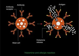
Histamine allergic reaction. Diagram showing the process that leads to the release of histamine during an allergic reaction. Cells called mast cells (orange) mediate this response

Background imageCell Collection: Intestine wall, artwork

Intestine wall, artwork
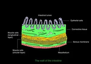
Intestine wall. Computer artwork of a section through the wall of a human intestine, showing its structure. For the labelled diagram see image: C009/1711


For sale as Licensed Images

Choose your image, Select your licence and Download the media

"Exploring the Intricacies of Life: Unveiling the Wonders of Cells" Delving into the microscopic world, a histological diagram of a mammalian retina reveals the intricate structure and organization of cells that enable us to perceive light. The cerebellum tissue, captured in a light micrograph, showcases its complex network of cells responsible for coordinating movement and balance. Intriguingly, even philosophers find solace in contemplating cellular existence. "Philosopher in Meditation, " an oil painting from 1632, reminds us that pondering life's mysteries often leads us back to our fundamental building blocks – cells. Zooming further into this realm, nerve and glial cells come alive under the lens. This captivating light micrograph highlights their vital role in transmitting signals throughout our nervous system. Meanwhile, a lavender pollen grain captured through scanning electron microscopy (SEM) displays nature's exquisite design at the cellular level. The battle against diseases takes center stage as T lymphocytes confront cancer cells in another SEM image. Science and evolution intertwine as we witness these tiny warriors fighting for survival within our bodies. Artistic expressions also shed light on cell significance; "The Death of Socrates, " painted in 1787, serves as a poignant reminder that even great minds are ultimately composed of countless individual cells. Santiago Ramon y Cajal's meticulous drawing from 1894 unveils various cell types within the mammalian cerebellum. His work not only represents scientific progress but also exemplifies how art can aid understanding by visually capturing complexity. Stepping away from biology momentarily, we encounter Rolls Royce/Snecma Olympus 593 Mk602 engine undergoing testing within a controlled environment called a test cell. Here too, precision engineering relies on understanding cellular mechanics to achieve optimal performance. Returning to biological marvels - HeLa cells take center stage under yet another microscope lens. These immortalized human cells have revolutionized medical research, paving the way for countless breakthroughs.