Cellular Collection (#9)

"Unveiling the Intricacies of Cellular Life: A Journey Through Micrographs" Budding yeast cell: Witness the birth of new life as a budding yeast cell emerges

Background imageCellular Collection: Chlamydomonas sp. algae, SEM

Chlamydomonas sp. algae, SEM
Green algae. Coloured scanning electron micrograph (SEM) of two Chlamydomonas sp. green algae. These single-celled organisms are aquatic, living in freshwater habitats

Background imageCellular Collection: Dividing cell, light micrograph

Dividing cell, light micrograph
Cell division. Fluorescent light micrograph of a cell that has divided by mitosis, the asexual replication of a cell into two new cells

Background imageCellular Collection: Liver cells, light micrograph

Liver cells, light micrograph
Liver cells. Light micrograph of a section through a human liver. The liver cells are pink with purple nuclei. A duct canal and its surrounding tissue can be seen at lower left

Background imageCellular Collection: Intestinal cells, light micrograph

Intestinal cells, light micrograph
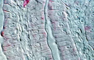
Intestinal cells. Light micrograph of a section through two folds (villi) in the small intestine. The villi are aligned vertically, with one at left and one at right

Background imageCellular Collection: Lung lining, SEM

Lung lining, SEM
Lung lining. Coloured scanning electron micrograph (SEM) of mucus-producing cells (orange, round) and cilia (yellow) lining a bronchus (lung airway)

Background imageCellular Collection: Large intestine, TEM

Large intestine, TEM
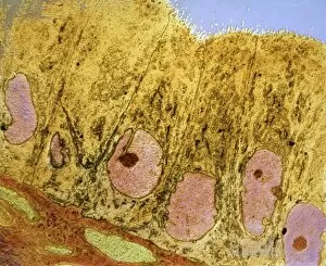
Large intestine. Coloured transmission electron micrograph (TEM) of a section through the wall of the large intestine. The surface consists of many small hair-like absorptive cells (green)

Background imageCellular Collection: Intestinal microvilli, SEM

Intestinal microvilli, SEM
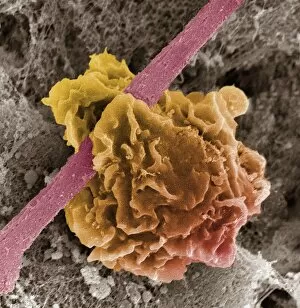
Intestinal microvilli. Coloured scanning electron micrograph (SEM) of microvilli from the duodenum, the first part of the small intestine

Background imageCellular Collection: Lung cells, light micrograph

Lung cells, light micrograph
Lung cells. Light micrograph of a section through the wall of a bronchus in the lungs. This is the upper layer of cells (the epithelium). The cell nuclei (round, dark) are across bottom

Background imageCellular Collection: Phagocytosis of a yeast spore, SEM

Phagocytosis of a yeast spore, SEM
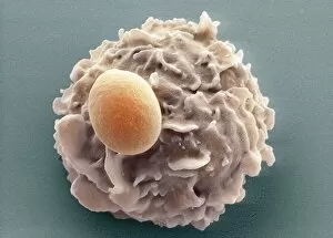
Phagocytosis of a yeast spore. Coloured scanning electron micrograph (SEM) of a neutrophil (brown) phagocytosing (engulfing and destroying) a spore from the yeast Candida albicans (orange)

Background imageCellular Collection: Dendritic cell, computer artwork

Dendritic cell, computer artwork
Dendritic cells. Computer artwork of dendritic cells, part of the human immune system. These are a type of macrophage cell found in the bodys tissues

Background imageCellular Collection: Phagocytosis of a fungus spore, SEM

Phagocytosis of a fungus spore, SEM
Phagocytosis of a fungus spore. Coloured scanning electron micrograph (SEM) of a spore from the fungus Aspergillus fumigatus (red) being phagocytosed (engulfed and destroyed)

Background imageCellular Collection: Dendritic cell, TEM

Dendritic cell, TEM
Dendritic cell. Coloured transmission electron micrograph (TEM) of a section through dendritic cells. These are part of the human immune system

Background imageCellular Collection: Man holding a mobile telephone to his ear

Man holding a mobile telephone to his ear
Mobile telephone. View of a man holding a mobile or cellular telephone to his ear. These telephones use low- power radio signals to transmit to a base station in the users local area, or cell

Background imageCellular Collection: Mobile phone rage

Mobile phone rage. Conceptual computer artwork of the head of an angry man poking from the screen of a mobile telephone. The use of these phones in public places has led to violent confrontations

Background imageCellular Collection: Mobile phone cartoon character

Mobile phone cartoon character

Background imageCellular Collection: Text message

Text message

Background imageCellular Collection: Mobile phones

Mobile phones
Mobile telephones, computer artwork

Background imageCellular Collection: Mobile phone X-ray

Mobile phone X-ray with a coloured map of the British Isles superimposed. This conceptual image could represent regional variation in patterns of mobile phone usage or network coverage

Background imageCellular Collection: Worldwide mobile telephone use

Worldwide mobile telephone use
Worldwide use of mobile (cellular) telephones, conceptual computer artwork

Background imageCellular Collection: Computer artwork of mans head with mobile phones

Computer artwork of mans head with mobile phones
Mobile telephone use. Computer illustration of a mans head seen in front of mobile, or cellular, telephones and brains. Mobile phones use low-power microwave radio signals to transmit calls

Background imageCellular Collection: WAP mobile telephone

WAP mobile telephone. Conceptual computer artwork of a hand holding a WAP-enabled mobile or cellular telephone. WAP (wireless application protocol)

Background imageCellular Collection: Avian flu virus, TEM

Avian flu virus, TEM
Avian flu. Coloured transmission electron micrograph (TEM) of influenza A virus particles (red). This is strain H5N1, which usually infects poultry, but began infecting humans in Hong Kong in 1997

Background imageCellular Collection: Water tower

Water tower with mobile telephone and television transmitting antennae. The water tower stores water for homes and businesses in the surrounding area

Background imageCellular Collection: Telecommunications mast

Telecommunications mast
Telecommunications base. Radio wave antennas mounted on top of a chimney. These are used to communicate between mobile phones in a telecommunications network

Background imageCellular Collection: Artwork of mobile telephone, globe & circuit board

Artwork of mobile telephone, globe & circuit board
Global telecommunication. Computer artwork of a hand holding a mobile cellular telephone over a globe and a circuit board, depicting global telecommunication

Background imageCellular Collection: Uterus cells, TEM

Uterus cells, TEM
Uterus cells. Coloured transmission electron micrograph (TEM) of endometrial cells (yellow) from the uterus. These cells form the endometrium, the epithelial tissue lining inside the uterus

Background imageCellular Collection: Salivary gland, light micrograph

Salivary gland, light micrograph
Salivary gland. Coloured light micrograph of a section through a sublingual salivary gland, which is situated directly under the tongue

Background imageCellular Collection: Macrophage ingesting debris, SEM

Macrophage ingesting debris, SEM
Macrophage ingesting cell debris at the site of a wound, coloured scanning electron micrograph (SEM). Macrophages are a type of large white blood cell that are part of the bodys immune system

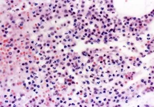
Background imageCellular Collection: Human blood cells, light micrograph

Human blood cells, light micrograph
Human blood cells. Light micrograph of blood cells at a site of inflammation, showing red blood cells (erythrocytes, red) surrounded by many granulocytes (blue)

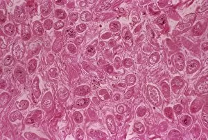
Background imageCellular Collection: Bilharzia infection, ureter tissue

Bilharzia infection, ureter tissue
Bilharzia infection. Light micrograph of human ureter tissue that contains numerous eggs from Schistosoma flukes. The ureters are tubes that carry urine from the kidneys to the bladder

Background imageCellular Collection: Rheumatoid arthritis mechanism, artwork

Rheumatoid arthritis mechanism, artwork
Rheumatoid arthritis mechanism. Artwork showing various aspects of rheumatoid arthritis, the inflammation of the joints due to attack by the bodys own immune system

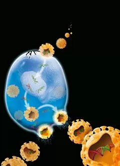
Background imageCellular Collection: Virus replication cycle, artwork

Virus replication cycle, artwork. The cycle starts at upper centre, as virus particles (orange) enter a cell (blue). Once in the cell the viral genetic material, deoxyribonucleic acid (DNA, green)

Background imageCellular Collection: Langerhans cell, TEM

Langerhans cell, TEM
Langerhans cell. Coloured transmission electron micrograph (TEM) of a section through a Langerhans cell from the epidermis of the skin. The tennis racket shaped objects are Birbeck granules

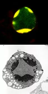
Background imageCellular Collection: Programmed cell death

Programmed cell death. Comparison of a fluorescent light micrograph (top) and transmission electron micrograph (TEM, bottom) of programmed cell death (apoptosis)

Background imageCellular Collection: Cultured cell, light micrograph

Cultured cell, light micrograph
Cultured cell. Immunofluorescent light micrograph of an epithelial cell (round, upper right). The cell proteins are marked by fluorescent dyes


For sale as Licensed Images

Choose your image, Select your licence and Download the media

"Unveiling the Intricacies of Cellular Life: A Journey Through Micrographs" Budding yeast cell: Witness the birth of new life as a budding yeast cell emerges, paving the way for future generations. Nerve and glial cells, light micrograph: Delve into the intricate network of nerve and glial cells that form the foundation of our nervous system. Dicotyledon plant stem, light micrograph: Explore the vibrant world within a dicotyledon plant stem, where cells work tirelessly to support growth and development. Rough endoplasmic reticulum, TEM: Peer into the fascinating realm of rough endoplasmic reticulum through a transmission electron microscope (TEM), revealing its role in protein synthesis. Dendritic cells, artwork: Marvel at an artistic representation showcasing dendritic cells' unique shape and function in immune responses. Cell structure: Uncover the hidden beauty within cellular structures as we unravel their complexity and significance in various biological processes. Mitosis, light micrograph: Observe nature's remarkable spectacle – mitosis – captured under a light microscope; witness how one cell divides into two with precision and grace. Cell division, fluorescent micrograph: Immerse yourself in a mesmerizing display of fluorescent colors capturing another facet of cell division's captivating dance. Dohle bodies in blood cell, micrograph: Discover peculiar "Dohle bodies" within blood cells through high-resolution microscopy—a clue to certain health conditions requiring further investigation. Acute promyelocytic leukemia, micrograph : Gain insight into acute promyelocytic leukemia by examining abnormal cellular characteristics observed under powerful magnification—an invaluable tool for diagnosis and treatment strategies Phagocytosis of fungal spores SEM : Witness nature's defense mechanism unfold as fungal spores are engulfed by cells in a captivating scanning electron microscope (SEM) image.