Cellular Collection (#6)

"Unveiling the Intricacies of Cellular Life: A Journey Through Micrographs" Budding yeast cell: Witness the birth of new life as a budding yeast cell emerges

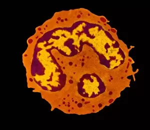
Background imageCellular Collection: Granulocyte white blood cell, TEM

Granulocyte white blood cell, TEM
Granulocyte white blood cell. Coloured transmission electron micrograph (TEM) of a neutrophil granulocyte, a type of white blood cell

Background imageCellular Collection: Acute leukaemia, light micrograph

Acute leukaemia, light micrograph
Acute leukaemia. Light micrograph of blood cells from bone marrow in a case of acute leukaemia. These cells include a polynuclear degranulated basophil and myeloblasts

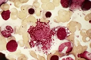
Background imageCellular Collection: Chronic myeloid leukaemia, micrograph

Chronic myeloid leukaemia, micrograph
Chronic myeloid leukaemia. Light micrograph of blood cells from bone marrow in a case of chronic myeloid leukaemia. Leukaemia is a cancer where certain blood cells form in excess

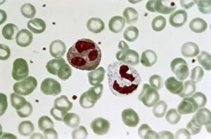
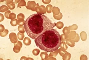
Background imageCellular Collection: Granulocyte blood cells, light micrograph

Granulocyte blood cells, light micrograph
Granulocyte blood cells. Light micrograph of two types of granulocytes, a type of white blood cell (leucocyte). At left is a neutrophil, with an eosinophil at right

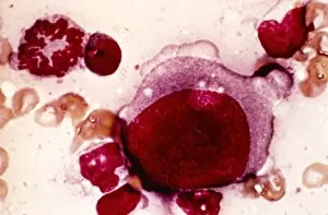
Background imageCellular Collection: Reticulosarcoma, light micrograph

Reticulosarcoma, light micrograph
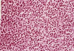
Reticulosarcoma. Light micrograph of reticulocyte blood cells (red, one at upper left) from a lymphatic ganglion in a case of reticulosarcoma

Background imageCellular Collection: Langerhans cell histiocytosis, micrograph

Langerhans cell histiocytosis, micrograph
Langerhans cell histiocytosis. Light micrograph of blood cells in a case of Langerhans cell histiocytosis (LCH), also called X-type histiocytosis

Background imageCellular Collection: Cell destruction, artwork

Cell destruction, artwork
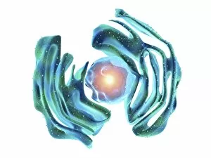
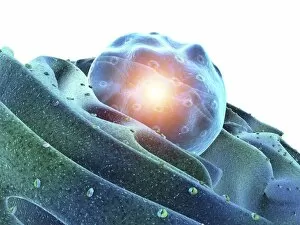
Cell destruction. Artwork of a cell undergoing destruction. This image could represent programmed cell death (apoptosis), a normal part of a cell life cycle

Background imageCellular Collection: Hodgkins disease, light micrograph

Hodgkins disease, light micrograph
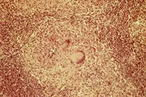
Hodgkins disease. Light micrograph of two non-dystrophic histiocyte blood cells (centre) in a case of Hodgkins disease, also called Hodgkins lymphoma

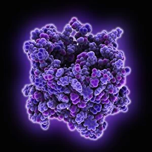
Background imageCellular Collection: VDAC-1 ion channel protein C015 / 8250

VDAC-1 ion channel protein C015 / 8250
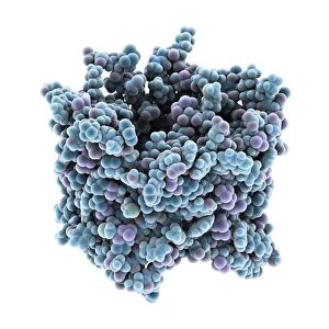
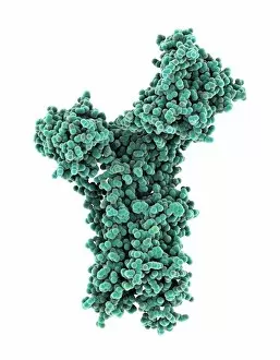
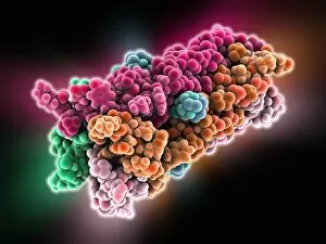
VDAC-1 ion channel protein, molecular model. This is the human voltage-dependent anion-selective channel protein 1 (VDAC-1)

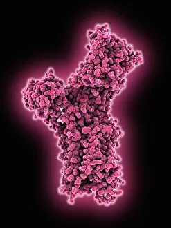
Background imageCellular Collection: Calcium ATPase ion pump C015 / 7143

Calcium ATPase ion pump C015 / 7143
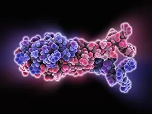
Calcium ATPase ion pump, molecular model. This enzyme is found in muscle cell membranes, where it pumps calcium in and out of muscle cells and controls muscle contractions

Background imageCellular Collection: Sodium-potassium ion pump proteins C015 / 9993

Sodium-potassium ion pump proteins C015 / 9993
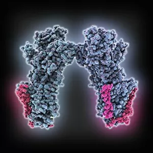
Sodium-potassium ion pump proteins, molecular model. Sodium-potassium ATPase (adenosine triphosphatase) is an ATP-powered ion pump found in all animal cells

Background imageCellular Collection: Sodium-potassium ion pump proteins C015 / 9997

Sodium-potassium ion pump proteins C015 / 9997
Sodium-potassium ion pump proteins, molecular model. Sodium-potassium ATPase (adenosine triphosphatase) is an ATP-powered ion pump found in all animal cells

Background imageCellular Collection: Reticulosarcoma, light micrograph C015 / 7133

Reticulosarcoma, light micrograph C015 / 7133
Reticulosarcoma. Light micrograph of reticulocyte blood cells (red, one at upper left) from a lymphatic ganglion in a case of reticulosarcoma

Background imageCellular Collection: VDAC-1 ion channel protein C015 / 8249

VDAC-1 ion channel protein C015 / 8249
VDAC-1 ion channel protein, molecular model. This is the human voltage-dependent anion-selective channel protein 1 (VDAC-1)

Background imageCellular Collection: Blood cells, light micrograph C015 / 7130

Blood cells, light micrograph C015 / 7130
Blood cells. Light micrograph of red blood cells (erythrocytes, red) and white blood cells (leucocytes, nuclei stained pink)

Background imageCellular Collection: Calcium ATPase ion pump C015 / 7142

Calcium ATPase ion pump C015 / 7142
Calcium ATPase ion pump, molecular model. This enzyme is found in muscle cell membranes, where it pumps calcium in and out of muscle cells and controls muscle contractions

Background imageCellular Collection: Cellular polarity, illustration C018 / 0775

Cellular polarity, illustration C018 / 0775
Cellular Polarity. Conceptual illustration of cell polarity research and its medical implications

Background imageCellular Collection: Ricin molecule, artwork C017 / 3656

Ricin molecule, artwork C017 / 3656
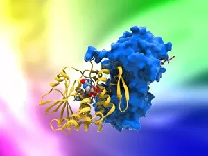
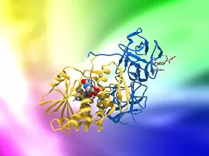
Ricin molecule. Computer artwork showing the structure of a molecule of the toxic protein ricin. Ricin comprises two entwined amino acid chains; A (yellow) and B (blue)

Background imageCellular Collection: Ricin molecule, artwork C017 / 3655

Ricin molecule, artwork C017 / 3655
Ricin molecule. Computer artwork showing the structure of a molecule of the toxic protein ricin. Ricin comprises two entwined amino acid chains; A (yellow) and B (blue)

Background imageCellular Collection: Ricin molecule, artwork C017 / 3648

Ricin molecule, artwork C017 / 3648
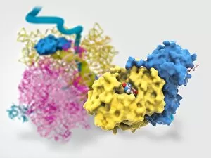
Ricin molecule Computer artwork showing the structure of a molecule of the toxic protein ricin (blue and yellow) with an active ribosome in the background

Background imageCellular Collection: Genetic metaphase analysis C019 / 0289

Genetic metaphase analysis C019 / 0289
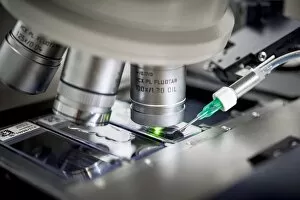
Genetic metaphase analysis. Close-up of a microscope being used in a genetics laboratory to analyse the metaphase stage of cell division

Background imageCellular Collection: Genetic metaphase analysis C019 / 0288

Genetic metaphase analysis C019 / 0288
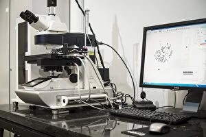
Genetic metaphase analysis. Equipment used in a genetics laboratory to analyse the metaphase stage of cell division. During this stage

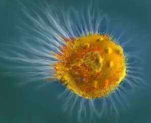
Background imageCellular Collection: Paramecium sp. protozoa (SEM) C014 / 2148

Paramecium sp. protozoa (SEM) C014 / 2148
Paramecium protozoa. Coloured scanning electron micrograph (SEM) of Paramecium sp. protozoa. These single-celled organisms are aquatic, being found in freshwater habitats

Background imageCellular Collection: ATP synthase molecule

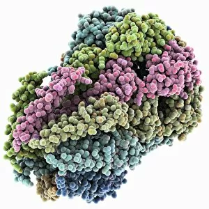
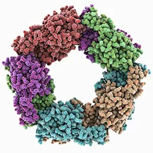
ATP synthase molecule. Molecular model showing the structure of ATP synthase (ATPase) subunit C. ATPase is an important enzyme that provides energy for cells through the synthesis of adenosine

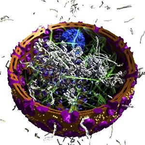
Background imageCellular Collection: Animal cell organelles, artwork

Animal cell organelles, artwork
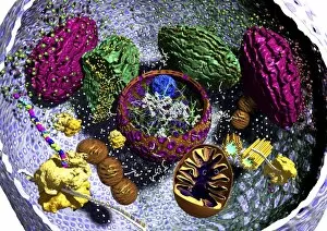
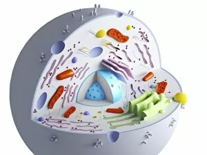
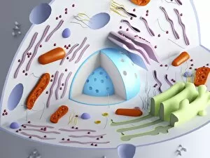
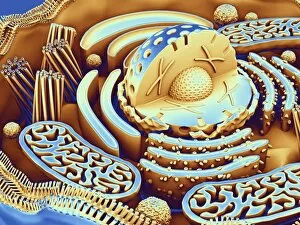
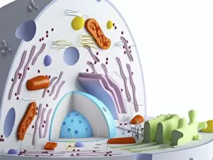
Animal cell organelles. Artwork showing the organelles in a eukaryotic cell. This is an animal cell. Structures include the nucleus (centre) which has a membrane with nuclear pores (purple)

Background imageCellular Collection: Human muscle fibres, diagram

Human muscle fibres, diagram
Human muscle fibres. Diagram showing the structure of human muscles from the cellular to the macroscopic level. At lower right, muscle cells contain many myofibrils

Background imageCellular Collection: MscS ion channel protein structure

MscS ion channel protein structure. Molecular model showing the protein structure of a Mechanosensitive Channel of Small Conductance (MscS) from an Escherichia coli bacterium

Background imageCellular Collection: Apple iPhone 4 mobile telephone

Apple iPhone 4 mobile telephone
Apple iPhone 4. The iPhone 4 mobile phone was introduced on 24 June 2010. It is a smart phone, having the ability to run various applications (known as apps)

Background imageCellular Collection: Eukaryotic cell nucleus, artwork

Eukaryotic cell nucleus, artwork
Eukaryotic cell nucleus. Artwork of the internal structure and contents of the nucleus of a eukaryotic cell. The nucleus has been sectioned in half

Background imageCellular Collection: Influenza proton pump, molecular model

Influenza proton pump, molecular model
Influenza proton pump. Molecular model showing the protein structure of a proton pump from an influenza virus. Proton pumps are membrane proteins that move protons across a cell membrane

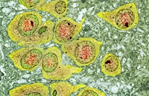
Background imageCellular Collection: Brain nerve cells, TEM C014 / 0356

Brain nerve cells, TEM C014 / 0356
Brain nerve cells. Transmission electron micrograph (TEM) of a section through brain tissue from the cerebral cortex, showing numerous neurons (nerve cells) surrounded by axons and dendrites

Background imageCellular Collection: H-NS chromatin-structuring protein

H-NS chromatin-structuring protein. Molecular model of the oligomerization domain of the H-NS protein from the Escherichia coli bacterium. This dimeric molecule folds in on itself, as shown here

Background imageCellular Collection: Chloride ion channel, molecular model

Chloride ion channel, molecular model. This is a ClC ion channel. Its role is to mediate the flow of chloride ions across cell membranes

Background imageCellular Collection: Potassium ion channel

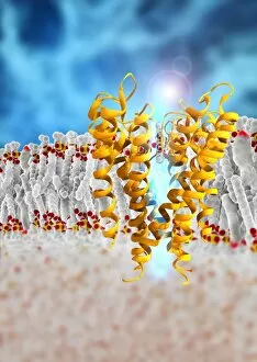
Potassium ion channel. Computer artwork of a KcsA potassium ion (K+) channel (ribbons) embedded in a phospholipid (spheres) cell membrane (horizontal, centre)

Background imageCellular Collection: Voltage-gated potassium channel

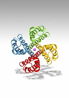
Voltage-gated potassium channel. Computer model showing the molecular structure of a voltage-gated potassium (Kv) ion channel

Background imageCellular Collection: Endometrial hyperplasia, light micrograph C015 / 7104

Endometrial hyperplasia, light micrograph C015 / 7104
Endometrial hyperplasia. Light micrograph of a section through tissue from the endometrium, in a case of endometrial hyperplasia. The endometrium is the uterus lining

Background imageCellular Collection: Sir3 gene silencer acting on DNA C015 / 7062

Sir3 gene silencer acting on DNA C015 / 7062
Sir3 gene silencer acting on DNA, molecular model. Sir3 (purple and grey) is acting on a circular strand of DNA (deoxyribonucleic acid, red)

Background imageCellular Collection: Sir3 gene silencer acting on DNA C015 / 7061

Sir3 gene silencer acting on DNA C015 / 7061
Sir3 gene silencer acting on DNA

Background imageCellular Collection: TFAM transcription factor bound to DNA C015 / 7060

TFAM transcription factor bound to DNA C015 / 7060
TFAM transcription factor bound to DNA, molecular model. Human mitochondrial transcription factor A (TFAM, pink) bound to a strand of DNA (deoxyribonucleic acid, orange and green)

Background imageCellular Collection: Peroxiredoxin 4 antioxidant enzyme C015 / 7022

Peroxiredoxin 4 antioxidant enzyme C015 / 7022
Peroxiredoxin 4 antioxidant enzyme, molecular model. This enzyme, also called peroxiredoxin IV (PrxIV), plays a catalytic role in cell metabolism on the endoplasmic reticulum

Background imageCellular Collection: Nucleus and endoplasmic reticulum C015 / 6797

Nucleus and endoplasmic reticulum C015 / 6797
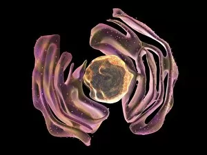
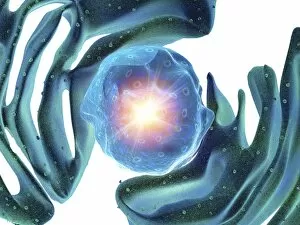
Computer artwork showing part of a human or eukaryotic cell. In the middle the nucleus which has a membrane with nuclear pores. Inside the nucleus is the DNA

Background imageCellular Collection: Animal cell organelles, artwork C015 / 6793

Animal cell organelles, artwork C015 / 6793
Animal cell organelles. Computer artwork showing the organelles in a eukaryotic cell. This is an animal cell. Structures include the nucleus (centre) which has a membrane with nuclear pores

Background imageCellular Collection: Animal cell organelles, artwork C015 / 6792

Animal cell organelles, artwork C015 / 6792
Animal cell organelles. Computer artwork showing the organelles in a eukaryotic cell. This is an animal cell. Structures include the nucleus (centre) which has a membrane with nuclear pores

Background imageCellular Collection: Nucleus and endoplasmic reticulum C015 / 6783

Nucleus and endoplasmic reticulum C015 / 6783
Computer artwork showing part of a human or eukaryotic cell. In the middle the nucleus which has a membrane with nuclear pores. Inside the nucleus is the DNA

Background imageCellular Collection: Nucleus and endoplasmic reticulum C015 / 6782

Nucleus and endoplasmic reticulum C015 / 6782
Computer artwork showing part of a human or eukaryotic cell. In the middle the nucleus which has a membrane with nuclear pores. Inside the nucleus is the DNA

Background imageCellular Collection: Nucleus and endoplasmic reticulum C015 / 6781

Nucleus and endoplasmic reticulum C015 / 6781
Computer artwork showing part of a human or eukaryotic cell. In the middle the nucleus which has a membrane with nuclear pores. Inside the nucleus is the DNA

Background imageCellular Collection: Animal cell organelles, artwork C015 / 6775

Animal cell organelles, artwork C015 / 6775
Animal cell organelles. Computer artwork showing the organelles in a eukaryotic cell. This is an animal cell. Structures include the nucleus (centre) which has a membrane with nuclear pores

Background imageCellular Collection: Animal cell organelles, artwork C015 / 6778

Animal cell organelles, artwork C015 / 6778
Animal cell organelles. Computer artwork showing the organelles in a eukaryotic cell. This is an animal cell. Structures include the nucleus (centre) which has a membrane with nuclear pores


For sale as Licensed Images

Choose your image, Select your licence and Download the media

"Unveiling the Intricacies of Cellular Life: A Journey Through Micrographs" Budding yeast cell: Witness the birth of new life as a budding yeast cell emerges, paving the way for future generations. Nerve and glial cells, light micrograph: Delve into the intricate network of nerve and glial cells that form the foundation of our nervous system. Dicotyledon plant stem, light micrograph: Explore the vibrant world within a dicotyledon plant stem, where cells work tirelessly to support growth and development. Rough endoplasmic reticulum, TEM: Peer into the fascinating realm of rough endoplasmic reticulum through a transmission electron microscope (TEM), revealing its role in protein synthesis. Dendritic cells, artwork: Marvel at an artistic representation showcasing dendritic cells' unique shape and function in immune responses. Cell structure: Uncover the hidden beauty within cellular structures as we unravel their complexity and significance in various biological processes. Mitosis, light micrograph: Observe nature's remarkable spectacle – mitosis – captured under a light microscope; witness how one cell divides into two with precision and grace. Cell division, fluorescent micrograph: Immerse yourself in a mesmerizing display of fluorescent colors capturing another facet of cell division's captivating dance. Dohle bodies in blood cell, micrograph: Discover peculiar "Dohle bodies" within blood cells through high-resolution microscopy—a clue to certain health conditions requiring further investigation. Acute promyelocytic leukemia, micrograph : Gain insight into acute promyelocytic leukemia by examining abnormal cellular characteristics observed under powerful magnification—an invaluable tool for diagnosis and treatment strategies Phagocytosis of fungal spores SEM : Witness nature's defense mechanism unfold as fungal spores are engulfed by cells in a captivating scanning electron microscope (SEM) image.