
Chromosome Collection (page 2)
"Unlocking the Mysteries of Life: Exploring the World of Chromosomes" Witness the intricate dance of life as cells divide through mitosis


For sale as Licensed Images
Choose your image, Select your licence and Download the media
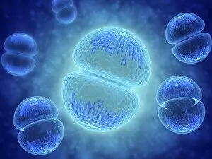
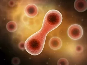
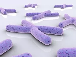
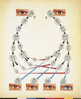
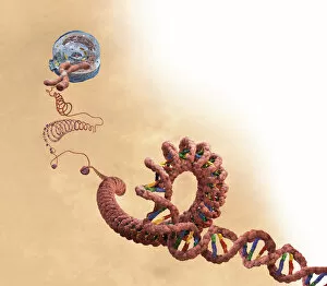
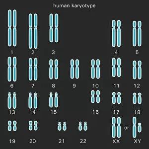
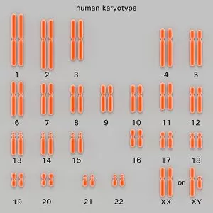
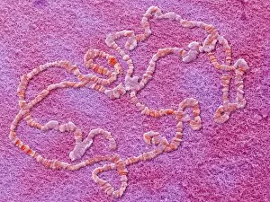
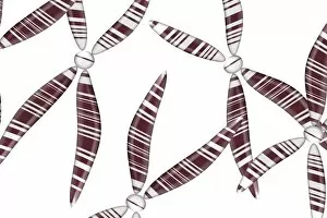
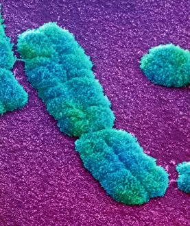
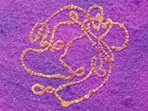
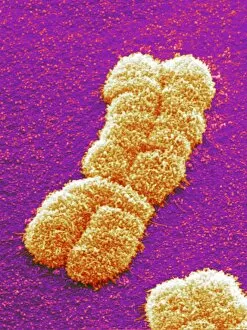
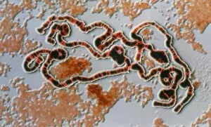
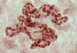
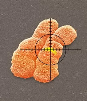
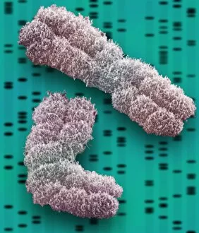
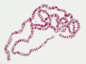
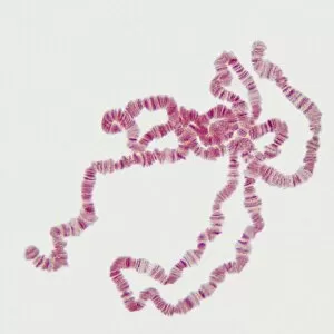
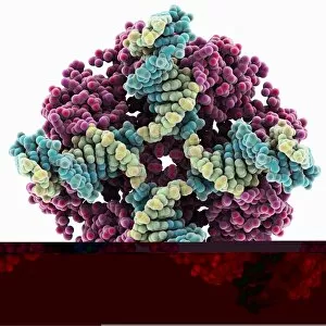
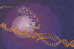
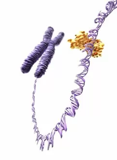
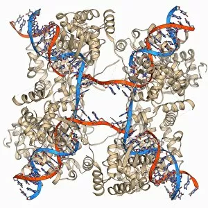
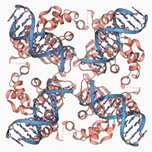
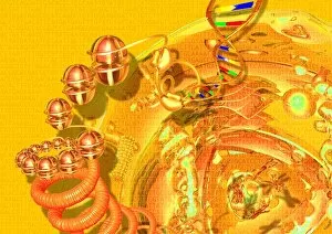
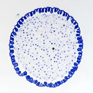
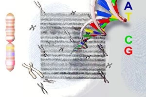
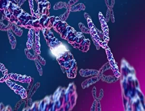
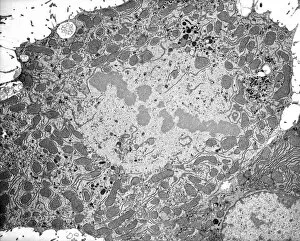
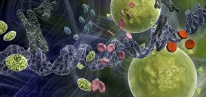
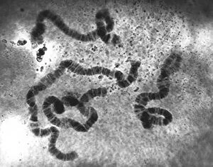
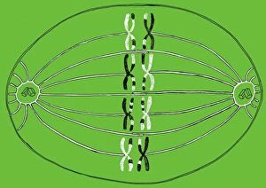
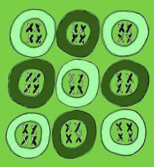
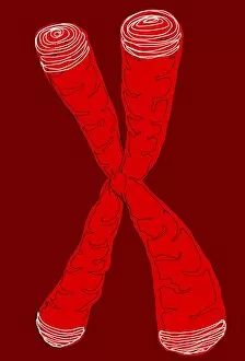
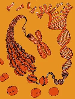
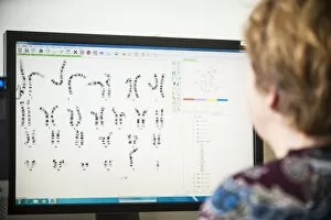
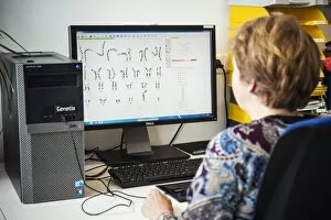
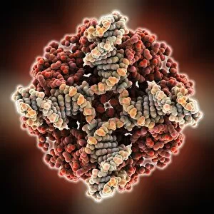
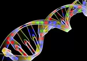
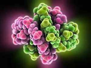
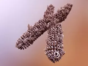
"Unlocking the Mysteries of Life: Exploring the World of Chromosomes" Witness the intricate dance of life as cells divide through mitosis, captured in a stunning light micrograph. Dive into the mesmerizing realm of cell division with a fluorescent micrograph, revealing the vibrant beauty hidden within. Behold a full set of male chromosomes under an SEM, offering a glimpse into our genetic blueprint and diversity. Marvel at the captivating sight of dividing cells, where new life emerges and growth takes place before your eyes. Delve deeper into cell division's enchantment with another fluorescent micrograph that showcases its breathtaking complexity. Unravel the secrets held within chromosome structure through an illuminating illustration that unveils their unique composition. Embark on a journey through DNA replication as bases attach to strands, forming two identical double DNA strands in this biomedical illustration. Explore protein synthesis and ribosome function in cross-section biomedical artwork that reveals how they contribute to cellular processes. Discover the essence of life encapsulated in each chromosome—a testament to our individuality and shared humanity. Traverse across nerve cells like astrocytes while contemplating their integral role in transmitting information throughout our complex nervous system. Immerse yourself in these captivating images—such as human chromosomes observed under an SEM—and let them ignite your curiosity about the wonders concealed within every living organism's DNA structure.