Chromosome Collection (#5)

"Unlocking the Mysteries of Life: Exploring the World of Chromosomes" Witness the intricate dance of life as cells divide through mitosis

Background imageChromosome Collection: Meiosis, SEM

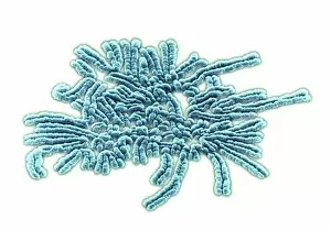
Meiosis, SEM
Meiosis. Coloured scanning electron micrograph (SEM) of chromosomes during metaphase (I) of meiosis (gamete formation). Chromosomes consist of deoxyribonucleic acid (DNA) and proteins

Background imageChromosome Collection: Computer-enhanced LM of human chromosomes

Computer-enhanced LM of human chromosomes
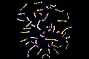
Chromosomes. Computer-enhanced light micrograph of human chromosomes. Chromosomes are thread-like structures in the cell nucleus that carry genetic information in the form of genes

Background imageChromosome Collection: Human chromosome 3

Human chromosome 3, computer artwork. Humans have 23 pairs of chromosomes in total. Chromosomes are made up of DNA (Deoxyribonucleic acid), which carries the bodys genetic information

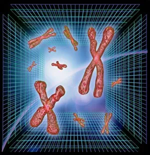
Background imageChromosome Collection: Computer artwork of chromosomes over grids

Computer artwork of chromosomes over grids

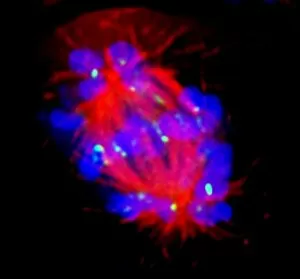
Background imageChromosome Collection: Abnormal mitosis

Abnormal mitosis
Mitosis. Fluorescence micrograph of a cell during abnormal anaphase of mitosis (nuclear division). During mitosis two daughter nuclei are formed from one parent nucleus

Background imageChromosome Collection: Dividing cell

Dividing cell. Differential interference contrast (DIC) light micrograph of a cell (lower left) in the metaphase stage of mitosis (cell division). The cells nuclei are stained with fluorescent dye

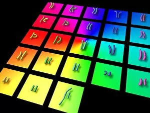
Background imageChromosome Collection: Male chromosome set

Male chromosome set
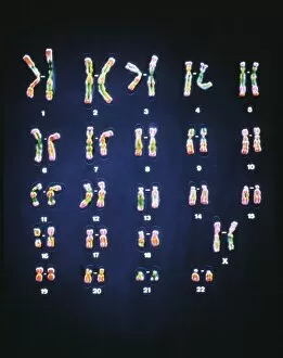
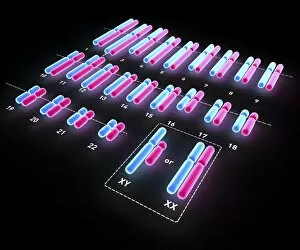
Male human chromosome set. Computer artwork of the complete set (karyotype) of healthy male human chromosomes. Humans have 46 chromosomes in most cells

Background imageChromosome Collection: Mitotic cell division, TEM

Mitotic cell division, TEM
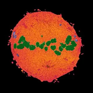
Mitotic cell division. Coloured transmission electron micrograph (TEM) of a cell during the metaphase stage of mitosis (cell division)

Background imageChromosome Collection: Gender determination

Gender determination, computer artwork. Gender is determined by the sex chromosomes, X and Y. Females have 2 X chromosomes and men an X and a Y chromosome

Background imageChromosome Collection: Male sex chromosomes

Male sex chromosomes. Computer artwork of an X and Y symbolising the male sex chromosomes. An individuals sex is determined by the presence or absence of the Y chromosome

Background imageChromosome Collection: Mitosis, TEM

Mitosis, TEM
Mitosis, coloured transmission electron micrograph (TEM). Longitudinal section through a human T cell in metaphase. During mitosis two daughter nuclei are formed from one parent nucleus

Background imageChromosome Collection: Cell mitosis

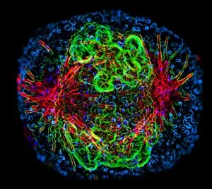
Cell mitosis. Confocal fluorescence light micrograph composite showing 6 stages of mitotic cell division. At far left, the cell has completed the first stage of cell division known as interphase in

Background imageChromosome Collection: Mitosis

Mitosis. Fluorescence micrograph of a cell during prometaphase of mitosis (nuclear division). During mitosis two daughter nuclei are formed from one parent nucleus

Background imageChromosome Collection: Chromosome, SEM

Chromosome, SEM
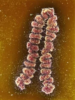
Chromosome. Coloured scanning electron micrograph (SEM) of a condensed chromosome showing chromomeres. Chromosomes, which consist of two identical chromatids joined at a centromere

Background imageChromosome Collection: Normal female chromosomes

Normal female chromosomes
False-colour light micrograph of a normal trypsin/leishman banded female karyotype, the full complement of female chromosomes arranged in numbered homologous pairs

Background imageChromosome Collection: Human chromosomes

Human chromosomes
False-colour light micrograph of a normal set of human chromosomes, the strands of DNA which lie within the nucleus of each human cell and carry the genetic code

Background imageChromosome Collection: Sperm fertilising an egg, artwork

Sperm fertilising an egg, artwork
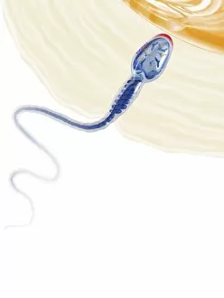
Sperm fertilising an egg. Cutaway artwork of a human sperm cell (spermatozoon) penetrating an egg cells (ovum) thick outer layer (zona pellucida). The egg cells membrane is at top right

Background imageChromosome Collection: Genetic modification

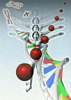
Genetic modification, conceptual artwork. Human figures and DNA (deoxyribonucleic acid, upper left to bottom right corner), which carries the genetic code. Chromosomes are seen at top left corner

Background imageChromosome Collection: Mitosis, artwork

Mitosis, artwork

Background imageChromosome Collection: X chromosome

X chromosome
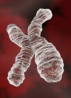
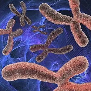
3D-computer illustration of a human X chromosome, one of the two sex chromosomes. Within almost every human cell are 46 chromosomes of which 22 pairs are the same in females and males

Background imageChromosome Collection: DNA construction, artwork

DNA construction, artwork
Conceptional computer artwork of a DNA being constructed or repaired

Background imageChromosome Collection: Genetic research, conceptual artwork

Genetic research, conceptual artwork
Genetic research, conceptual computer artwork

Background imageChromosome Collection: Chromosome analysis

Chromosome analysis
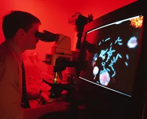
MODEL RELEASED. Chromosome analysis. Geneticist analysing human chromosomes by fluorescent microscopy. Individual chromosomes are visible on the computer monitor as pale blue rods

Background imageChromosome Collection: Human chromosomes, artwork

Human chromosomes, artwork
Human karyotype, computer artwork. A karyotype is the complete set of chromosomes of an organism. Humans have 46 chromosomes in total

Background imageChromosome Collection: Artwork of bacterial DNA: chromosome and plasmids

Artwork of bacterial DNA: chromosome and plasmids

Background imageChromosome Collection: Human karyotype, artwork

Human karyotype, artwork
Human karyotype, computer artwork. A karyotype is the complete set of chromosomes of an organism. Humans have 46 chromosomes in total

Background imageChromosome Collection: Chromosomes and DNA

Chromosomes and DNA (deoxyribonucleic acid) molecules, computer artwork. Chromosomes are composed of DNA coiled around proteins

Background imageChromosome Collection: Chromosomes

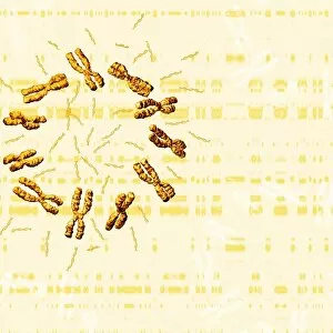
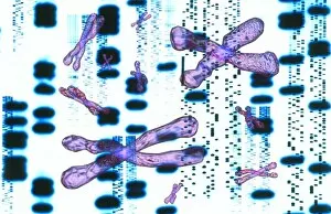
Chromosomes and DNA (deoxyribonucleic acid) molecules on a DNA autoradiogram, computer artwork. Chromosomes are composed of deoxyribonucleic acid (DNA) coiled around proteins

Background imageChromosome Collection: DNA

DNA
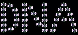
Deoxyribonucleic acid. Conceptual artwork of DNA (deoxyribonucleic acid) spelt out with fluoresence micrographs of anaphase stage cells. Anaphase is a stage of mitosis (nuclear division)

Background imageChromosome Collection: Gene switch, conceptual artwork

Gene switch, conceptual artwork
Gene switch. Conceptual computer artwork representing a gene switch, showing a switch (red) on one of the arms of a chromosome

Background imageChromosome Collection: Artwork of DNA sequences and chromosomes

Artwork of DNA sequences and chromosomes
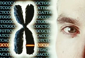
DNA sequences & chromosomes. Computer artwork of an autoradiogram of DNA (deoxyribonucleic acid) sequences. A close up view of a sequence is superimposed in the foreground

Background imageChromosome Collection: Eye colour genetics

Eye colour genetics


For sale as Licensed Images

Choose your image, Select your licence and Download the media

"Unlocking the Mysteries of Life: Exploring the World of Chromosomes" Witness the intricate dance of life as cells divide through mitosis, captured in a stunning light micrograph. Dive into the mesmerizing realm of cell division with a fluorescent micrograph, revealing the vibrant beauty hidden within. Behold a full set of male chromosomes under an SEM, offering a glimpse into our genetic blueprint and diversity. Marvel at the captivating sight of dividing cells, where new life emerges and growth takes place before your eyes. Delve deeper into cell division's enchantment with another fluorescent micrograph that showcases its breathtaking complexity. Unravel the secrets held within chromosome structure through an illuminating illustration that unveils their unique composition. Embark on a journey through DNA replication as bases attach to strands, forming two identical double DNA strands in this biomedical illustration. Explore protein synthesis and ribosome function in cross-section biomedical artwork that reveals how they contribute to cellular processes. Discover the essence of life encapsulated in each chromosome—a testament to our individuality and shared humanity. Traverse across nerve cells like astrocytes while contemplating their integral role in transmitting information throughout our complex nervous system. Immerse yourself in these captivating images—such as human chromosomes observed under an SEM—and let them ignite your curiosity about the wonders concealed within every living organism's DNA structure.