
Clotted Collection
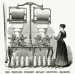
In 1875, the Tompkins upright rotary knitting machine revolutionized the textile industry with its innovative design


For sale as Licensed Images
Choose your image, Select your licence and Download the media
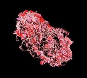
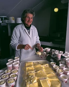
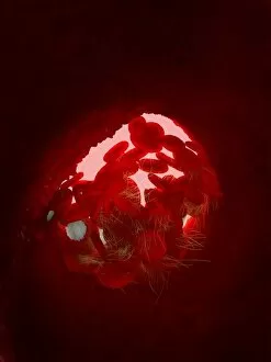
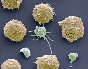
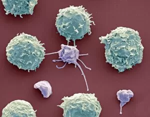
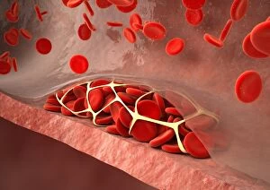
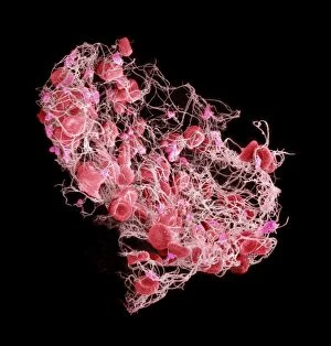
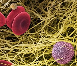
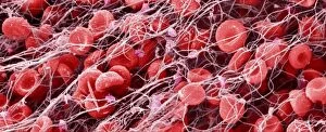
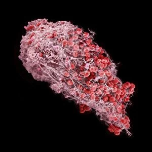
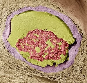
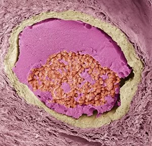
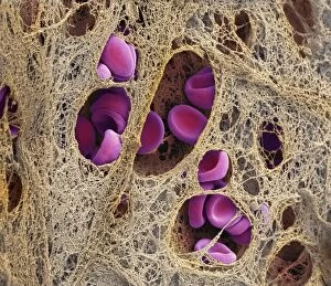
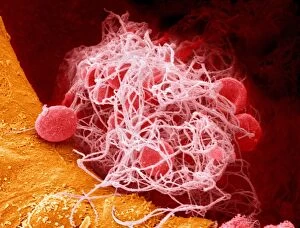
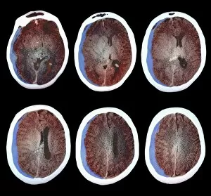
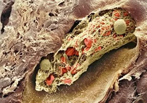
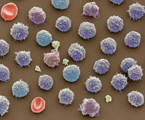
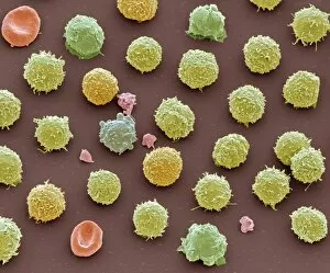
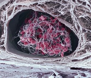
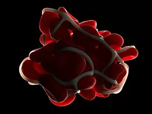
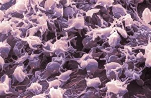
In 1875, the Tompkins upright rotary knitting machine revolutionized the textile industry with its innovative design. Little did anyone know that a similar concept would occur within our own bodies - the formation of blood clots. A captivating SEM image (C016 / 9747) showcases the intricate structure of a blood clot, resembling an abstract work of art. Just like a skilled server delicately serving Roskillys Cornish Clotted Cream, our body's natural defense mechanism works tirelessly to prevent excessive bleeding and promote healing. However, sometimes this process can go awry. Ischaemia, as depicted in a digital angiogram, highlights how blocked arteries can lead to serious health complications. The artwork (C013 / 4649) depicting thrombosed blood vessels serves as a stark reminder of the potential dangers lurking within our circulatory system. But amidst these medical marvels lies beauty in unexpected places. White blood cells and platelets captured under SEM (C016 / 3098 & C016 / 3099) resemble ethereal landscapes or celestial formations - nature's way of protecting us from harm. As we drag a spoon through thick white cream in a bowl, it mirrors the mesmerizing flow and texture reminiscent of clotting blood (SEM C016 / 9751 & C016 / 9746). It reminds us that even something seemingly mundane can hold profound significance when viewed through different lenses. So next time you encounter "clotted, " whether it be on your dessert or in scientific research images, take a moment to appreciate both its literal and metaphorical implications. From groundbreaking inventions to life-saving mechanisms within our bodies – clotted is not just about cream; it represents resilience, protection, and the delicate balance between life and death.