
Collagen Collection (#2)
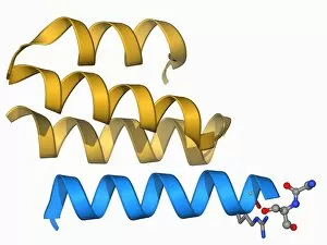
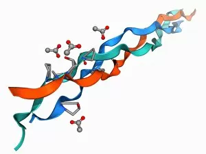
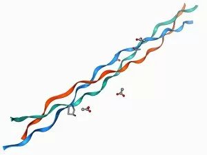
Collagen, the building block of our body's connective tissues, plays a crucial role in various biological processes


For sale as Licensed Images
Choose your image, Select your licence and Download the media
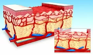
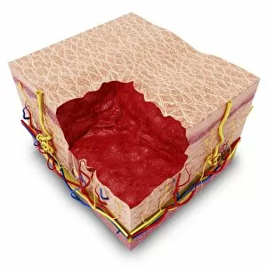
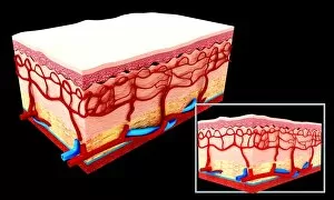
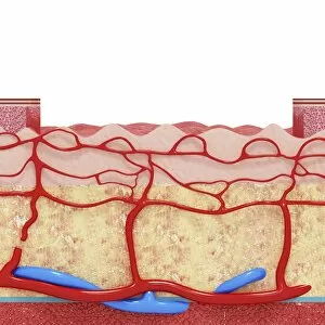
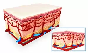
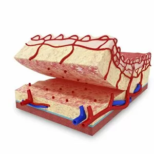
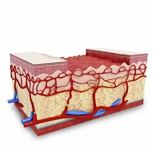
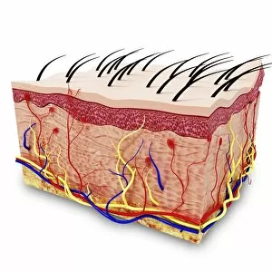
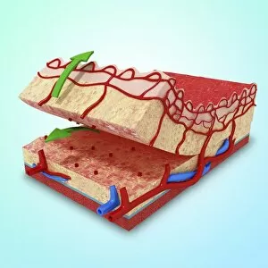
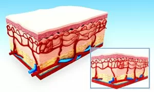
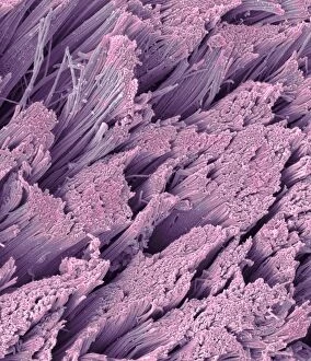
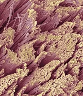
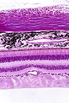
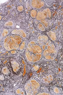
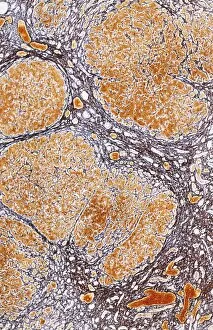
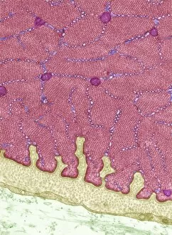
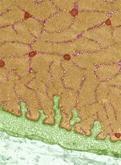
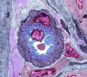
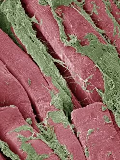
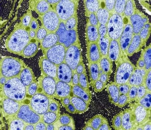
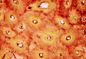
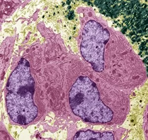
Collagen, the building block of our body's connective tissues, plays a crucial role in various biological processes. Under the scanning electron microscope (SEM), collagen fibers appear as intricate networks, resembling delicate artwork (C016 / 9873). One significant function is its involvement in the blood coagulation cascade. It forms a mesh-like structure that aids in clot formation and prevents excessive bleeding. This process can be visualized through captivating illustrations (Collagen synthesis and assembly). In tendons, collagen fibers provide strength and flexibility to support movement. SEM images reveal their organized arrangement, highlighting their importance for maintaining structural integrity. Within eye muscles, transmission electron microscopy (TEM) captures the detailed architecture fibers (C014 / 1468). These fibers contribute to the precise movements required for proper vision. Beyond these microscopic wonders lies a macroscopic world where collagen impacts bone health. In whale bone tissue captured under light micrography, we witness how this protein contributes to the strength and resilience of these majestic creatures. Illustrations depicting human bone formation showcase how collagen scaffolds serve as foundations for healthy bones' growth and development. However, when osteoporosis strikes, osteoclasts take center stage by breaking down bone tissue relentlessly. These destructive osteoclasts are contrasted with another group of cells called osteoblasts who diligently work towards building healthy bones by depositing new layers of collagen-rich matrix. Sadly though, in individuals suffering from osteoporosis or weakened bones due to aging or other factors; osteoclasts erode away more bone than is rebuilt by osteoblasts leading to fragile skeletal structures susceptible to fractures. Understanding the multifaceted roles played by collagen allows us to appreciate its significance not only on a cellular level but also within our bodies as it supports vital functions like blood clotting and maintains strong bones essential for overall well-being.