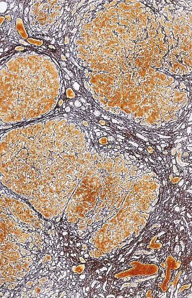
Cirrhosis of liver, light micrograph C016 / 0529
![]()

Wall Art and Photo Gifts from Science Photo Library
Cirrhosis of liver, light micrograph C016 / 0529
Cirrhosis of liver. Light micrograph of a section through liver tissue, damaged by cirrhosis. Cirrhosis occurs as a result of a range of factors causing damage to liver function. The disease typically destroys hepatocyte liver cells and the normal morphology of the liver lobules. Conditions leading to cirrhosis include chronic alcoholism, hepatitis and obstruction of bile flow. Because the hepatocytes are naturally regenerative, agents that cause their death stimulate new hepatocyte growth seen here as oval-shaped nodules. A further feature of cirrhosis is excessive build up of fibrous tissue, resulting from over-production of collagen fibres. This all means the cirrhotic liver is resistant to blood flowing through it and therefore cannot perform it s
Science Photo Library features Science and Medical images including photos and illustrations
Media ID 9238259
© MICROSCAPE/SCIENCE PHOTO LIBRARY
Cell Biology Cirrhosis Collagen Cytological Cytology Diseased Fibre Fibres Fibrous Hepatic Hepatocyte Hepatocytes Histological Histology Liver Stain Stained Tissue Abnormal Cells Condition Disorder Light Micrograph Light Microscope Section Sectioned Unhealthy
EDITORS COMMENTS
This print from Science Photo Library showcases the intricate details of cirrhosis of the liver at a microscopic level. Cirrhosis, a condition characterized by extensive damage to liver function, is caused by various factors such as chronic alcoholism, hepatitis, and bile flow obstruction. In this light micrograph, we observe the detrimental effects of cirrhosis on liver tissue. The disease destroys hepatocyte liver cells and disrupts the normal structure of liver lobules. However, due to their regenerative nature, new oval-shaped nodules can be seen here as evidence of hepatocyte growth stimulated by agents causing cell death. Another prominent feature depicted in this image is the excessive accumulation of fibrous tissue resulting from over-production of collagen fibers. This fibrosis further hampers blood flow through the cirrhotic liver and impairs its ability to perform vital functions. The detailed anatomy captured in this photograph provides valuable insights into the unhealthy state of an organ affected by cirrhosis. It serves as a reminder that conditions leading to this disorder can have severe consequences for human health. Microscape/Science Photo Library has expertly captured both the biological and histological aspects of cirrhosis in this visually striking image print.
MADE IN THE USA
Safe Shipping with 30 Day Money Back Guarantee
FREE PERSONALISATION*
We are proud to offer a range of customisation features including Personalised Captions, Color Filters and Picture Zoom Tools
SECURE PAYMENTS
We happily accept a wide range of payment options so you can pay for the things you need in the way that is most convenient for you
* Options may vary by product and licensing agreement. Zoomed Pictures can be adjusted in the Cart.

