
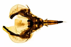
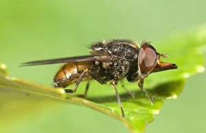
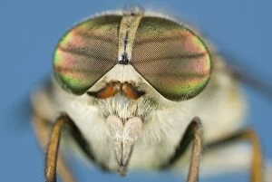
Compound Eye Collection (#8)
The compound eye, a marvel of nature's engineering, is a fascinating feature found in various insects


For sale as Licensed Images
Choose your image, Select your licence and Download the media
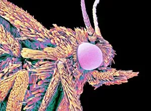
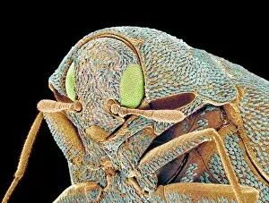
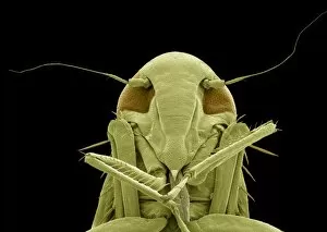
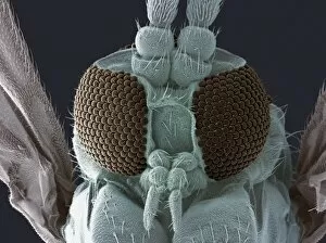
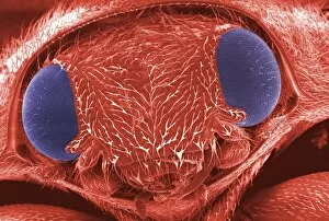
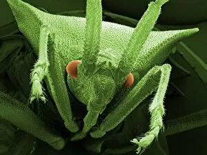
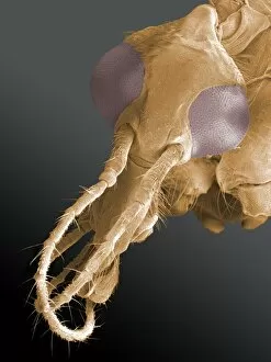
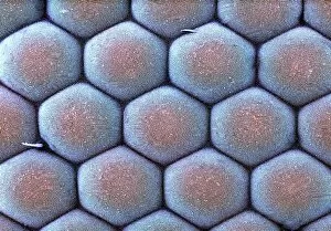
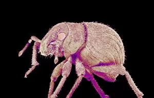
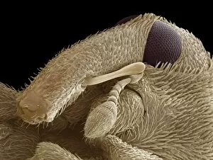
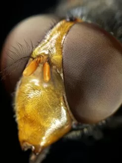
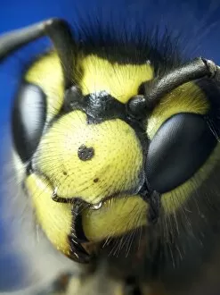
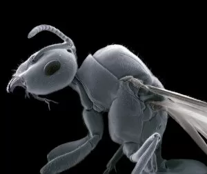
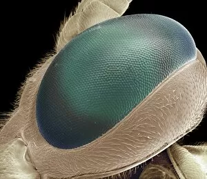
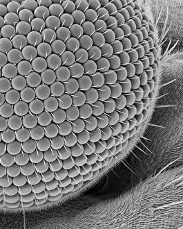
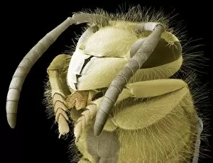
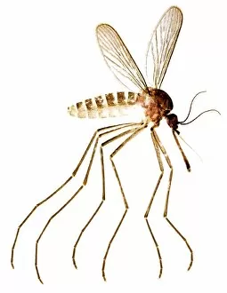
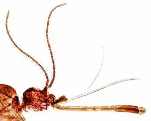
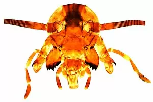
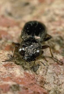
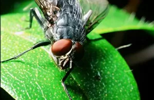
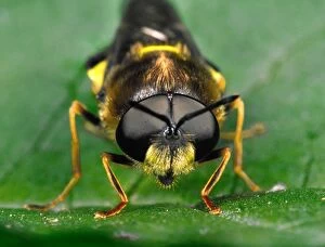
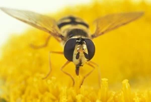
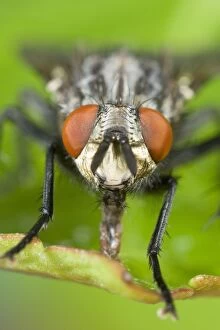
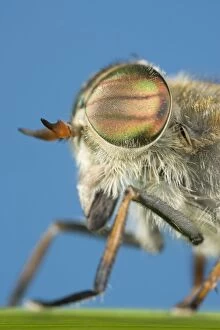
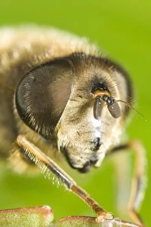
The compound eye, a marvel of nature's engineering, is a fascinating feature found in various insects. Take the fruit fly for instance, as seen through the lens of an SEM Z340 / 0768. Magnified at x300 on an A4 size frame, its compound eye reveals intricate details that captivate our imagination. But it's not just the fruit fly that showcases this incredible structure. The Culex mosquito and red-barbed ant also possess compound eyes that have been meticulously examined under scanning electron micrographs (SEM). These images provide us with glimpses into their world, where vision takes on a whole new dimension. Moving on to the head of a honey bee captured by another SEM image, we witness how these tiny creatures rely heavily on their compound eyes for navigation and finding nectar-rich flowers amidst vast landscapes, and is truly remarkable how such small organisms can possess such complex visual systems. And let's not forget about other species like the hornet mimic hoverfly or even mosquitoes themselves. Their internal anatomy has been revealed through cross-sections and SEM images showcasing their feeding habits - including blood-sucking from human skin. Zooming back in to focus solely on flies, we encounter yet another stunning view of a house fly's compound eye magnified at x40 using SEM technology. This close-up view allows us to appreciate the intricate arrangement of individual lenses called ommatidia which make up this unique visual organ. With beekeeping being an essential practice worldwide, understanding the compound eye becomes crucial in managing beehives effectively. By comprehending how bees perceive their surroundings through these specialized organs, beekeepers can ensure optimal conditions for honey production and colony health. Finally returning to our initial subject - the fruit fly - we delve deeper into its microscopic world with another SEM image (Z340 / 0699).