Cornea Collection (#3)

The cornea, a remarkable part of the eye anatomy, is often depicted in stunning artwork

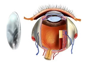
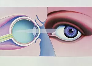
Background imageCornea Collection: Contact lens and eye anatomy, artwork C016 / 6572

Contact lens and eye anatomy, artwork C016 / 6572
Contact lens and eye anatomy. Artwork of a contact lens (left) and the anatomy of an eye (right) with a contact lens placed on the corneal surface (upper right)

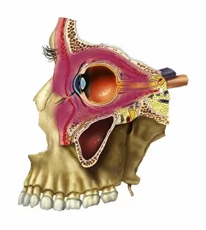
Background imageCornea Collection: Plegmon eye inflammation, artwork

Plegmon eye inflammation, artwork
Plegmon eye inflammation. Artwork of a sagittal section through a human eye and the surrounding structures, in a case of a subcutaneous (below the skin) phlegmon inflammation of an eye cavity

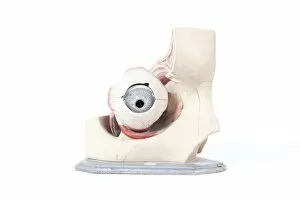
Background imageCornea Collection: Human eye, historical anatomical model

Human eye, historical anatomical model
Human eye. 1940s model showing the anatomy of the human eye

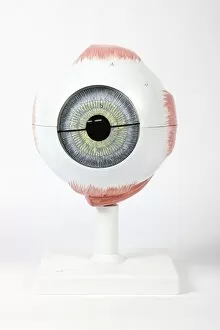
Background imageCornea Collection: Human eye, anatomical model

Human eye, anatomical model
Eye anatomy. Anatomical model showing the structure of the human eye

Background imageCornea Collection: Human eyes, anatomical models

Human eyes, anatomical models

Background imageCornea Collection: Eye anatomy, artwork C016 / 8742

Eye anatomy, artwork C016 / 8742
Eye anatomy, artwork. The eye is seen in cutaway format from the side. Structures shown include the lens (light blue, lower left), its attachment points, the iris

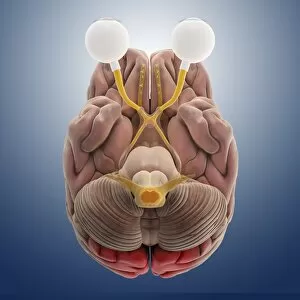
Background imageCornea Collection: Brain and eye anatomy, artwork C013 / 4664

Brain and eye anatomy, artwork C013 / 4664
Brain and eye anatomy. Computer artwork of the brain from below, with the front of the brain and the eyeballs (white, one at right sectioned) at top

Background imageCornea Collection: Artwork of human eye in front view and in section

Artwork of human eye in front view and in section

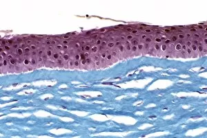
Background imageCornea Collection: Cornea

Cornea. Light micrograph of a section through a cornea, the transparent region at the front of the eye. The outer surface of the cornea is a stratified epithelium (dark pink)

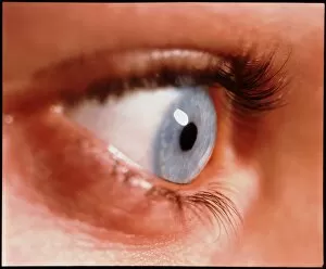
Background imageCornea Collection: Close-up of a womans healthy blue eye

Close-up of a womans healthy blue eye

Background imageCornea Collection: Cornea, SEM

Cornea, SEM
Cornea. Coloured scanning electron micrograph (SEM) of a section through a cornea, the transparent region on the outer surface of the eye. The front of the eye is at top

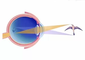
Background imageCornea Collection: Artwork of eye in section demonstrating vision

Artwork of eye in section demonstrating vision
Longsightedness. Artwork of a section through the human eye demonstrating longsightedness. In longsightedness or hyperopia close objects are seen blurred

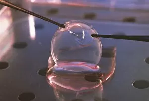
Background imageCornea Collection: Corneal strip

Corneal strip grown in the laboratory. It has been cultured from human epithelial cells that line the cornea, the outer shell of the eyeball

Background imageCornea Collection: Glaucoma

Glaucoma. Artwork showing the flow of the watery aqueous humour (arrows) in a human eye with glaucoma, abnormally high pressure in the eye

Background imageCornea Collection: Shortsightedness

Shortsightedness. Artwork of section through an eyeball affected by shortsightedness, or myopia. This is a condition in which distant objects appear blurred

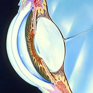
Background imageCornea Collection: Artwork of the structure of the front of an eye

Artwork of the structure of the front of an eye

Background imageCornea Collection: Developing pig eye, light micrograph

Developing pig eye, light micrograph. From right the structures seen are: the cornea (brown), the lens (purple), the retina (pink), the choroid (dark brown line) and the sclera (white of the eye)

Background imageCornea Collection: Bird eye anatomy, artwork

Bird eye anatomy, artwork. The structure of a birds eye is similar to that of humans and other vertebrates, with a clear cornea forming a bulge (left) in front of the lens (oval)

Background imageCornea Collection: Eye lens and accommodation, diagram

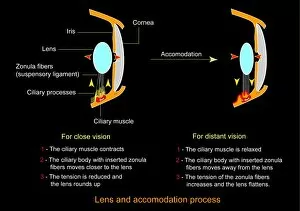
Eye lens and accommodation, diagram
Eye lens and accommodation. Diagram showing the process by which the eye changes the shape of the lens when changing between close vision and distant vision. This process is known as accommodation

Background imageCornea Collection: Eye lens and accommodation, artwork

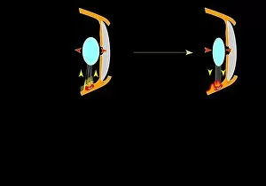
Eye lens and accommodation, artwork
Eye lens and accommodation. Artwork showing the process by which the eye changes the shape of the lens when changing between close vision and distant vision. This process is known as accommodation

Background imageCornea Collection: Dissected eye, 1844 artwork

Dissected eye, 1844 artwork
Dissected eye. This anatomical artwork is figure 1, plate 78 from volume 3 (1844) of Traite complet de l anatomie de l homme (1831-1854)

Background imageCornea Collection: Eye anatomy, 1844 artwork

Eye anatomy, 1844 artwork
Eye anatomy, front view. This anatomical artwork is figure 2, plate 75 from volume 3 (1844) of Traite complet de l anatomie de l homme (1831-1854)

Background imageCornea Collection: Human eyeball, artwork

Human eyeball, artwork
Human eyeball. Artwork of a human eyeball, showing the outer sclera (white) and the structures at the front and back. At the front, the eye bulges in a protrusion called the cornea

Background imageCornea Collection: Eye laser therapy, artwork

Eye laser therapy, artwork
Eye laser therapy. Artwork showing three stages in laser therapy to correct corneal deformation. The outer layer of the cornea is cut open and a flap (left) pulled back

Background imageCornea Collection: Eye anatomy

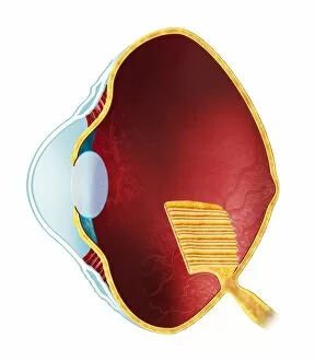
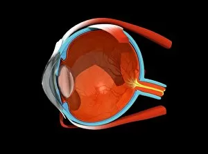
Eye anatomy. Cutaway artwork passing vertically through a human eye, showing its internal anatomy and structure. The front of the eye is at left, and the structures here include the cornea

Background imageCornea Collection: Biometric eye scan, artwork

Biometric eye scan, artwork
Biometric eye scan, computer artwork. Eye being scanned for identifying features (such as patterns in the iris) that can be used in security systems to restrict access to authorised individuals

Background imageCornea Collection: Keratan sulphate, molecular model

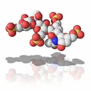
Keratan sulphate, molecular model. This linear polysaccharide polymer is found in cartilage, bone and the cornea (transparent part of the eye)

Background imageCornea Collection: Dogfish head, transverse section

Dogfish head, transverse section
Dogfish head. Light micrograph of a transverse section through the head of a lesser spotted dogfish (Scyliorhinus canicula), in the region of the eyes (upper left and upper right)

Background imageCornea Collection: Rabbit eye, longitudinal section

Rabbit eye, longitudinal section
Rabbit eye. Light micrograph of a longitudinal section through the eye of a rabbit (Oryctolagus cuniculus). The rabbit has a typical mammalian eye structure


For sale as Licensed Images

Choose your image, Select your licence and Download the media

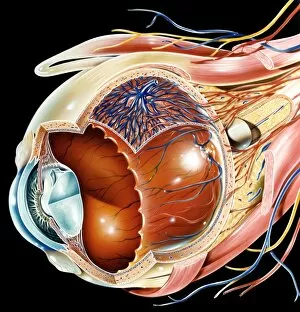
The cornea, a remarkable part of the eye anatomy, is often depicted in stunning artwork. From digital illustrations showcasing the dilation and contraction of outer radial muscle fibers to conceptual artworks representing laser eye surgery, this vital component never fails to captivate our imagination. In one captivating image, a human eye is beautifully illustrated with intricate details highlighting its external anatomy. Labels guide us through each structure, allowing us to appreciate the complexity and precision involved in vision. Another illustration takes us deeper into the normal anatomy of the eye through a cross-section view. This visual representation unveils the intricacies hidden beneath our eyelids and reminds us of the delicate balance required for optimal vision. A black-and-white photograph from Romania showcases an enchanting moment captured between a peasant woman and her cornea. The depth conveyed in this image speaks volumes about how crucial this small yet powerful organ is for connecting individuals with their surroundings. An ancient plate from Traite Complet de l'Anatomie de l'Homme offers insight into historical perspectives on eye surgery. This conceptual artwork transports us back in time, reminding us of humanity's relentless pursuit to understand and improve upon our own biology. Even nature itself provides inspiration when examining corneas; an engraving from The Pictorial Museum of Animated Nature features a common fly's head with its proboscis delicately resting against its transparent cornea. Such images remind us that even creatures so different from ourselves share similar anatomical wonders. Whether it be through scientific diagrams or artistic interpretations, exploring the beauty and complexity of corneas allows us to marvel at one of nature's most incredible creations – our eyesight itself.