
Crystalline Collection (#3)

"Captivating Crystalline Beauty

Background imageCrystalline Collection: Pepperming leaves (Mentha piperita), preparation for making medicine or liquor

Pepperming leaves (Mentha piperita), preparation for making medicine or liquor, on a rustic wooden underground

Background imageCrystalline Collection: Sage leaves (Salvia), preparation for making medicine or liquor, on a rustic wooden underground

Sage leaves (Salvia), preparation for making medicine or liquor, on a rustic wooden underground

Background imageCrystalline Collection: A bottle of hazelnut oil and some ripe and unripe hazelnuts (Corylus avellana)

A bottle of hazelnut oil and some ripe and unripe hazelnuts (Corylus avellana) on rustic wooden boards

Background imageCrystalline Collection: Artichoke on fine china in a luxurious ambience

Artichoke on fine china in a luxurious ambience

Background imageCrystalline Collection: Luxuriously set coffee table with silver coffee and tea pots, and a fruit pie on a silver cake plate

Luxuriously set coffee table with silver coffee and tea pots, and a fruit pie on a silver cake plate

Background imageCrystalline Collection: Stylish, colourful tinted champagne glasses on a silver platter

Stylish, colourful tinted champagne glasses on a silver platter

Background imageCrystalline Collection: Pizzette, small pizzas with buffalo mozzarella, chorizo and coriander, front

Pizzette, small pizzas with buffalo mozzarella, chorizo and coriander, front, and tomatoes and anchovies, rear

Background imageCrystalline Collection: Blurred street atmosphere through a window pane with rain drops

Blurred street atmosphere through a window pane with rain drops

Background imageCrystalline Collection: Rain drops on a window pane, blurry buildings in the back

Rain drops on a window pane, blurry buildings in the back

Background imageCrystalline Collection: Rain drops on a window pane, blurry buildings and cars in the back

Rain drops on a window pane, blurry buildings and cars in the back

Background imageCrystalline Collection: Olive oil

Olive oil

Background imageCrystalline Collection: Picture No. 11050178

Picture No. 11050178
Quartz Schist Dawson City, Yukon, Canada Date:

Background imageCrystalline Collection: Picture No. 11050177

Picture No. 11050177
Mica Schist Nevada, USA Date:

Background imageCrystalline Collection: Picture No. 11050176

Picture No. 11050176
Mica Schist Ketchikan, Alaska Date:

Background imageCrystalline Collection: Minerals, Pyromorphite (Lead chlorophosphate) green crystalline mineral on quartz

Minerals, Pyromorphite (Lead chlorophosphate) green crystalline mineral on quartz, from Mid-Wales orefield

Background imageCrystalline Collection: Picture No. 10734828

Picture No. 10734828
Crystalline Gold Specimen - 96 ounces Date:

Background imageCrystalline Collection: Picture No. 10870722

Picture No. 10870722
Western Australia - couple snorkelling in the crystalline waters of the lagoon of Ningaloo Reef Date:

Background imageCrystalline Collection: Ball and Stick Model showing arrangement of Carbon Atoms in Diamond

Ball and Stick Model showing arrangement of Carbon Atoms in Diamond

Background imageCrystalline Collection: Crystalline brucite in groundmass, close-up

Crystalline brucite in groundmass, close-up

Background imageCrystalline Collection: Ulexite in crystalline slab

Ulexite in crystalline slab

Background imageCrystalline Collection: Pile of fine sugar granules, view from above

Pile of fine sugar granules, view from above

Background imageCrystalline Collection: A clear salt grinder with rock salt scattered in front of it

A clear salt grinder with rock salt scattered in front of it

Background imageCrystalline Collection: Iron pyrite crystals, SEM

Iron pyrite crystals, SEM
Iron pyrite crystals, coloured scanning electron micrograph (SEM). Iron pyrite (iron disulphide, FeS2), commonly known as fools gold, is the most abundant sulphide mineral and often occurs as cubes

Background imageCrystalline Collection: Pesticide contamination, SEM

Pesticide contamination, SEM
Pesticide food contamination. Coloured scanning electron micrograph (SEM) of a single crystal of a fungicide on a broad bean plants leaf

Background imageCrystalline Collection: Iron pyrite crystal, SEM

Iron pyrite crystal, SEM
Iron pyrite crystal, coloured scanning electron micrograph (SEM). Iron pyrite (iron disulphide, FeS2), commonly known as fools gold, is the most abundant sulphide mineral and often occurs as cubes

Background imageCrystalline Collection: Phosphate crystals, SEM C014 / 1436

Phosphate crystals, SEM C014 / 1436
Phosphate crystals, coloured scanning electron micrograph (SEM). Magnification: x500 when printed at 10 centimetres wide

Background imageCrystalline Collection: Phosphate crystals, SEM C014 / 1435

Phosphate crystals, SEM C014 / 1435
Phosphate crystals, coloured scanning electron micrograph (SEM). Magnification: x500 when printed at 10 centimetres wide

Background imageCrystalline Collection: Muonionalusta meteorite, micrograph

Muonionalusta meteorite, micrograph
Dark field micrograph of the surfcace of the iron fine octahedrite type meteorite Muonionalusta, found in Sweden. The Widmanstatten pattern of lines on the newly-cut surface are produced by the edges

Background imageCrystalline Collection: Copper sulphate crystals light micrograph

Copper sulphate crystals light micrograph
Copper sulphate crystals, polarised light micrograph. Magnification: x2.5, when printed 10 centimetres wide

Background imageCrystalline Collection: Crushed diamond, SEM C016 / 9101

Crushed diamond, SEM C016 / 9101
Crushed diamond. Coloured scanning electron micrograph (SEM) of a crushed diamond crystal. Magnification: x250, when printed 10 centimetres wide

Background imageCrystalline Collection: Graphite, molecular model C016 / 8894

Graphite, molecular model C016 / 8894
Graphite, molecular model. Graphite is used in pencil leads and as a lubricant. It is composed of parallel layers of hexagonally arranged carbon atoms (spheres)

Background imageCrystalline Collection: Crushed diamond, SEM C016 / 9099

Crushed diamond, SEM C016 / 9099
Crushed diamond. Coloured scanning electron micrograph (SEM) of a crushed diamond crystal. Magnification: x250, when printed 10 centimetres wide

Background imageCrystalline Collection: Graphite, molecular model C016 / 8895

Graphite, molecular model C016 / 8895
Graphite, molecular model. Graphite is used in pencil leads and as a lubricant. It is composed of parallel layers of hexagonally arranged carbon atoms (spheres)

Background imageCrystalline Collection: Graphite, molecular model C016 / 8896

Graphite, molecular model C016 / 8896
Graphite, molecular model. Graphite is used in pencil leads and as a lubricant. It is composed of parallel layers of hexagonally arranged carbon atoms (spheres)

Background imageCrystalline Collection: Crushed diamond, SEM C016 / 9100

Crushed diamond, SEM C016 / 9100
Crushed diamond. Coloured scanning electron micrograph (SEM) of a crushed diamond crystal. Magnification: x250, when printed 10 centimetres wide

Background imageCrystalline Collection: Graphite, molecular model C016 / 8893

Graphite, molecular model C016 / 8893
Graphite, molecular model. Graphite is used in pencil leads and as a lubricant. It is composed of parallel layers of hexagonally arranged carbon atoms (spheres)

Background imageCrystalline Collection: Sodium chloride structure C016 / 8873

Sodium chloride structure C016 / 8873
Sodium chloride structure, molecular model. Sodium chloride (common salt) consists of a cubic lattice of sodium (small spheres) and chlorine (green) ions and has the chemical formula NaCl

Background imageCrystalline Collection: Crushed diamond, SEM C016 / 9102

Crushed diamond, SEM C016 / 9102
Crushed diamond. Coloured scanning electron micrograph (SEM) showing the surface of a crushed diamond. Magnification: x1600, when printed 10 centimetres wide

Background imageCrystalline Collection: Crushed diamond, SEM C016 / 9098

Crushed diamond, SEM C016 / 9098
Crushed diamond. Coloured scanning electron micrograph (SEM) showing the surface of a crushed diamond. Magnification: x1600, when printed 10 centimetres wide

Background imageCrystalline Collection: Tourmaline specimen C016 / 5991

Tourmaline specimen C016 / 5991
Tourmaline. A composite, doubly-terminated crystal of schorlite, with a little limonite and crystal of apatite. From Minerals of Cornwall and Devon (1987) by P.G. Embrey and R.F. Symes

Background imageCrystalline Collection: Baryte C016 / 6088

Baryte C016 / 6088
Baryte (barium sulphate). Specimen of the mineral Baryte from Wheal Mary Ann Menheriot, Cornwall, UK. Natural History Museum, London, UK

Background imageCrystalline Collection: Galena C016 / 6087

Galena C016 / 6087
Galena (lead sulphide). Specimen number BM 42141, from Heredsfoot Mine, Cornwall, UK. Natural History Museum, London, U

Background imageCrystalline Collection: Sugar types C015 / 8110

Sugar types C015 / 8110
Sugar types. The sugar types arrayed here are granulated sugar (piles at left), sugar cubes (pile at centre left), icing sugar (also called powdered sugar, piles at far right)

Background imageCrystalline Collection: Eye lens layers, SEM C018 / 0522

Eye lens layers, SEM C018 / 0522
Eye lens layers, coloured scanning electron micrograph (SEM). The lenses in mammalian eyes are made up of layers upon layers of tiny cells that interlock with one another via a series of tiny

Background imageCrystalline Collection: Eye lens layers, SEM C018 / 0523

Eye lens layers, SEM C018 / 0523
Eye lens layers, coloured scanning electron micrograph (SEM). The lenses in mammalian eyes are made up of layers upon layers of tiny cells that interlock with one another via a series of tiny

Background imageCrystalline Collection: Eye lens layers, SEM C018 / 0524

Eye lens layers, SEM C018 / 0524
Eye lens layers, coloured scanning electron micrograph (SEM). The lenses in mammalian eyes are made up of layers upon layers of tiny cells that interlock with one another via a series of tiny

Background imageCrystalline Collection: Phosphate crystal, SEM

Phosphate crystal, SEM
Coloured scanning electron micrograph (SEM) of a single phosphate crystal. A phosphate is an inorganic chemical and a salt of phosphoric acid

Background imageCrystalline Collection: Methyl sulphonal crystals, micrograph

Methyl sulphonal crystals, micrograph
Methyl sulphonal crystals, polarised light micrograph. Magnification: x114 when printed 10 centimetres wide


For sale as Licensed Images

Choose your image, Select your licence and Download the media

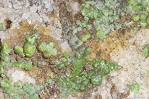
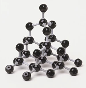
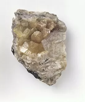
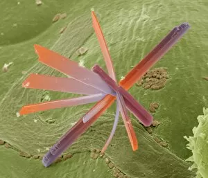
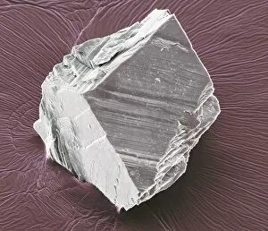
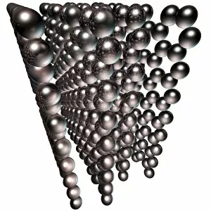
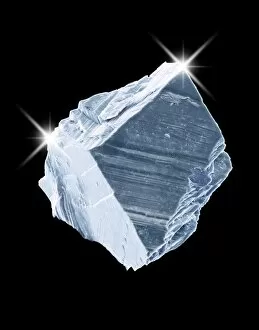
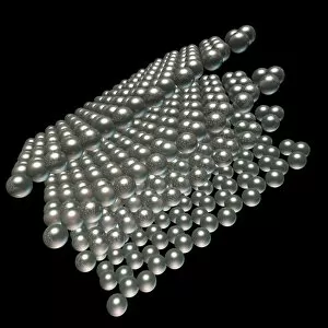
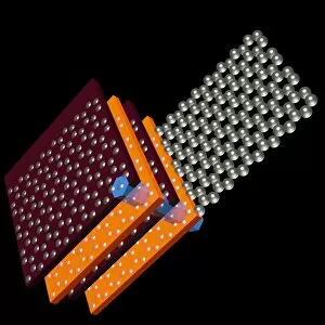
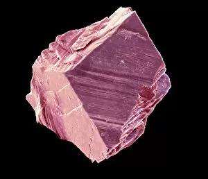
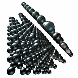
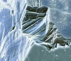
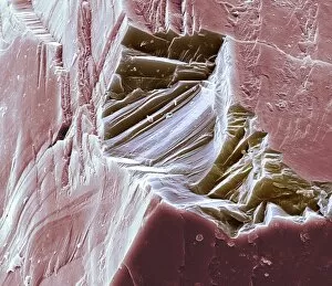
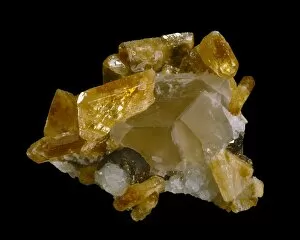
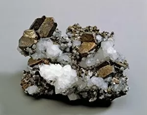
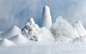
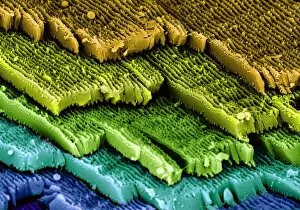
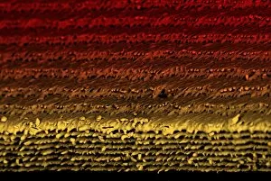
"Captivating Crystalline Beauty: A Glimpse into the Intricate World of Minerals and Molecules" Delicate copper and magnesium sulphate crystals under a powerful light microscope (LM) reveal their mesmerizing structure. Caffeine crystals, captured in a stunning light micrograph, showcase the intricate patterns formed by this stimulating compound. The perovskite crystal structure unveils its remarkable symmetry, creating an enchanting visual spectacle. A computer artwork depicts the brilliance of a diamond's crystalline lattice, reflecting its timeless allure. Cortisol crystals, magnified through a light micrograph (LM), exhibit their unique formation within our body's natural stress hormone. Vibrant copper sulphate crystals come to life when observed under a high-resolution light microscope (LM), revealing their captivating hues and shapes. The beach of Capo Coda Cavallo in Sardinia, Italy offers breathtaking views where nature's crystalline beauty meets pristine shores - truly an idyllic escape in Europe. Another glimpse into caffeine's microscopic world showcases its delicate crystal formations through an exquisite light micrograph. An artistic representation reveals the intricate patterns found within graphene sheets - highlighting both scientific marvels and aesthetic wonders. Baking soda crystals take on extraordinary shapes when examined using scanning electron microscopy (SEM), unveiling hidden textures that captivate the eye. Copper sulphate crystals shimmer with elegance as they are unveiled through the lens of a high-resolution light microscope (LM). A fly delicately perched on sugar granules is brought to life through scanning electron microscopy (SEM), showcasing how even everyday substances can possess unexpected beauty. In this collection of images and descriptions lies proof that there is unparalleled beauty hidden within every corner of our world – from minerals to molecules – reminding us to appreciate nature's artistry at both macroscopic and microscopic levels.