Cytological Collection (#7)

"Cytological Wonders Unveiled: Exploring the Intricacies of Cellular Life" In this captivating journey into the microscopic world, we delve into the realm of cytology

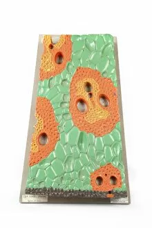
Background imageCytological Collection: Human tissue structure, historical model

Human tissue structure, historical model
Human tissue structure. 1960s anatomical model of a section through human body tissue, showing the arrangement and structure of the cells

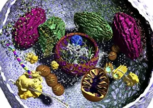
Background imageCytological Collection: Animal cell organelles, artwork

Animal cell organelles, artwork
Animal cell organelles. Artwork showing the organelles in a eukaryotic cell. This is an animal cell. Structures include the nucleus (centre) which has a membrane with nuclear pores (purple)

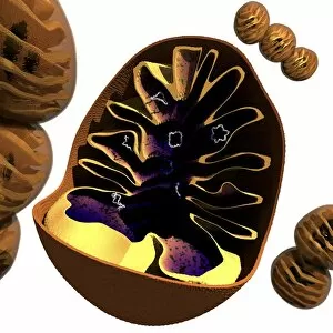
Background imageCytological Collection: Mitochondrion structure, artwork

Mitochondrion structure, artwork
Mitochondrion structure. Artwork showing the internal structure of a mitochondrion. This structure, found in eukaryotic cells, is the site of energy production

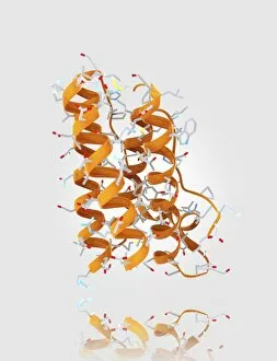
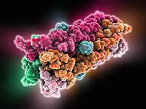
Background imageCytological Collection: Leptin molecule C014 / 4907

Leptin molecule C014 / 4907
Leptin molecule. Computer model showing the crystal structure of a molecule of the hormone leptin. Leptin is produced by adipose (fat) tissue

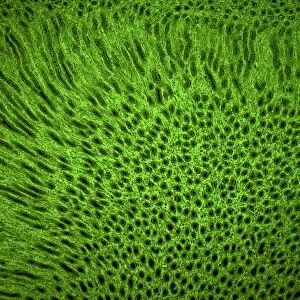
Background imageCytological Collection: Squamous epithelium C014 / 4643

Squamous epithelium C014 / 4643
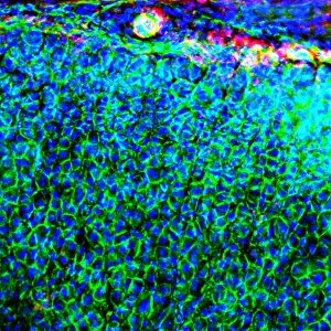
Squamous epithelium. Laser scanning confocal micrograph (LSCM) of a section through stratified squamous epithelial cells. Epithelial tissues line the cavities

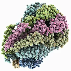
Background imageCytological Collection: MscS ion channel protein structure

MscS ion channel protein structure. Molecular model showing the protein structure of a Mechanosensitive Channel of Small Conductance (MscS) from an Escherichia coli bacterium

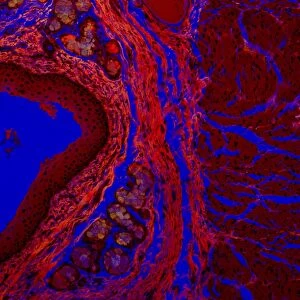
Background imageCytological Collection: Squamous epithelium, confocal micrograph C014 / 4642

Squamous epithelium, confocal micrograph C014 / 4642
Squamous epithelium. Laser scanning confocal micrograph (LSCM) of a section through stratified squamous epithelial cells. Epithelial tissues line the cavities

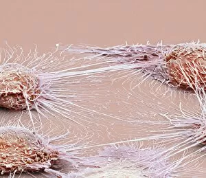
Background imageCytological Collection: Mouth cancer cells, SEM

Mouth cancer cells, SEM
Mouth cancer cells. Coloured scanning electron micrograph (SEM) of squamous cell carcinoma (cancer) cells from a human mouth

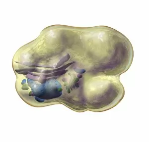
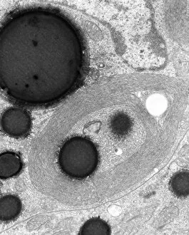
Background imageCytological Collection: Fat cell anatomy, artwork

Fat cell anatomy, artwork
Fat cell anatomy. Artwork of an adipocyte (fat cell) and its internal organelles. The majority of the cell is filled with a lipid (fat) droplet

Background imageCytological Collection: Eukaryotic cell nucleus, artwork

Eukaryotic cell nucleus, artwork
Eukaryotic cell nucleus. Artwork of the internal structure and contents of the nucleus of a eukaryotic cell. The nucleus has been sectioned in half

Background imageCytological Collection: Mitochondrial structure, artwork

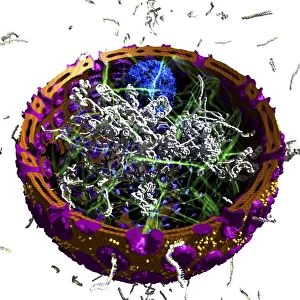
Mitochondrial structure, artwork
Mitochondrial structure. Computer artwork of a section through a mitochondrion, showing the internal structure and a loop of mitochondrial DNA (deoxyribonucleic acid, mtDNA)

Background imageCytological Collection: Leptin molecule and fat cells C014 / 4906

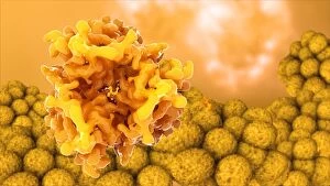
Leptin molecule and fat cells C014 / 4906
Leptin molecule and fat cells. Computer artwork showing a molecule of the hormone leptin with adipose (fat) cells (round), from which it is produced

Background imageCytological Collection: Mouth cancer cell dividing, SEM

Mouth cancer cell dividing, SEM
Mouth cancer dividing. Coloured scanning electron micrograph (SEM) of a squamous cell carcinoma (cancer) cell from a human mouth undergoing mitosis (nuclear division)

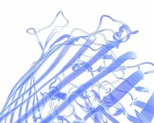
Background imageCytological Collection: Bacterial outer membrane protein molecule

Bacterial outer membrane protein molecule. Computer model showing a part of the secondary structure of a molecule of outer membrane protein A from Escherichia coli (E. coli) bacteria

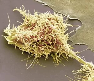
Background imageCytological Collection: Mouth cancer cell, SEM

Mouth cancer cell, SEM
Mouth cancer cell. Coloured scanning electron micrograph (SEM) showing the surface of a squamous cell carcinoma (cancer) cell from a human mouth

Background imageCytological Collection: Influenza proton pump, molecular model

Influenza proton pump, molecular model
Influenza proton pump. Molecular model showing the protein structure of a proton pump from an influenza virus. Proton pumps are membrane proteins that move protons across a cell membrane

Background imageCytological Collection: Arteriole, TEM

Arteriole, TEM
Arteriole. Transmission electron micrograph (TEM) of a section through a small calibre arterial vessel called an arteriole

Background imageCytological Collection: Smooth endoplasmic reticulum, TEM

Smooth endoplasmic reticulum, TEM
Smooth endoplasmic reticulum. Transmission electron micrograph (TEM) showing smooth endoplasmic reticulum (ER, thin lines) inside a cell that is synthesising steroid hormones

Background imageCytological Collection: Small bowel epithelium, TEM

Small bowel epithelium, TEM
Small bowel epithelium. Transmission electron micrograph (TEM) of a section through the epithelial lining of a villus in the small bowel, showing the enterocyte cells

Background imageCytological Collection: Optogenetics, conceptual artwork

Optogenetics, conceptual artwork. Optogenetics is the integration of optical and genetic systems to control precisely defined events within specific cells of living tissue

Background imageCytological Collection: Kidney tissue, confocal micrograph C014 / 4611

Kidney tissue, confocal micrograph C014 / 4611
Kidney tissue. Laser scanning confocal micrograph (LSCM) of a section through a human kidney, showing the cell nuclei (dark)

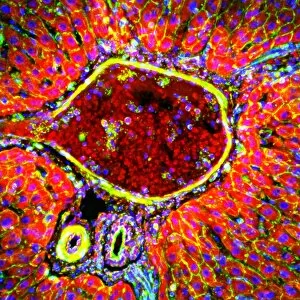
Background imageCytological Collection: Liver portal triad, light micrograph C016 / 8490

Liver portal triad, light micrograph C016 / 8490
Liver portal triad. Fluorescence deconvolution micrograph of a section through a portal triad in liver tissue, showing hepatocyte cells (red)

Background imageCytological Collection: Liver portal triad, light micrograph C016 / 8489

Liver portal triad, light micrograph C016 / 8489
Liver portal triad. Fluorescence deconvolution micrograph of a section through a portal triad in liver tissue, showing hepatocyte cells (red)

Background imageCytological Collection: Liver portal triad, light micrograph C016 / 8488

Liver portal triad, light micrograph C016 / 8488
Liver portal triad. Fluorescence deconvolution micrograph of a section through a portal triad in liver tissue, showing hepatocyte cells (red)

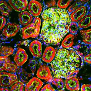
Background imageCytological Collection: Kidney tissue, fluorescence micrograph C016 / 8484

Kidney tissue, fluorescence micrograph C016 / 8484
Kidney tissue. Fluorescence deconvolution micrograph of a section through a kidney, showing glomeruli (green), cell nuclei (blue dots), and renal tubules (red, circular)

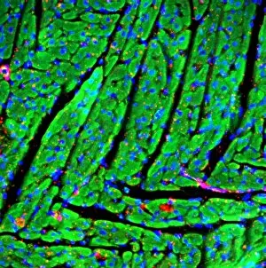
Background imageCytological Collection: Heart muscle, fluorescence micrograph C016 / 8483

Heart muscle, fluorescence micrograph C016 / 8483
Heart muscle. Fluorescence deconvolution micrograph of a section through heart tissue, showing the angular distribution of the myocardium (cardiac muscle) fibres (green)

Background imageCytological Collection: Heart muscle, fluorescence micrograph C016 / 8481

Heart muscle, fluorescence micrograph C016 / 8481
Heart muscle. Fluorescence deconvolution micrograph of a section through heart tissue, showing blood vessels (oval, centre-left and upper centre) running through the myocardium (cardiac muscle)

Background imageCytological Collection: Heart muscle, fluorescence micrograph C016 / 8478

Heart muscle, fluorescence micrograph C016 / 8478
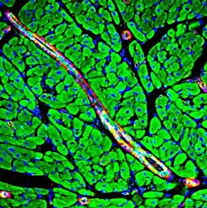
Heart muscle. Fluorescence deconvolution micrograph of a section through heart tissue, showing a blood vessel (diagonal, centre) running through the myocardium (cardiac muscle, green)

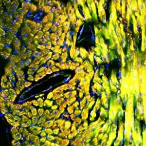
Background imageCytological Collection: Heart muscle, fluorescence micrograph C016 / 8477

Heart muscle, fluorescence micrograph C016 / 8477
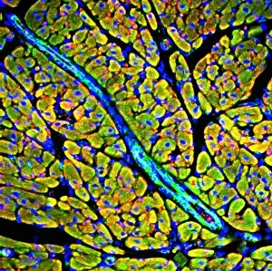
Heart muscle. Fluorescence deconvolution micrograph of a section through heart tissue, showing a blood vessel (blue, diagonal, centre) running through the myocardium (cardiac muscle, green)

Background imageCytological Collection: Adrenal gland, fluorescence micrograph C016 / 8472

Adrenal gland, fluorescence micrograph C016 / 8472
Adrenal gland. Fluorescence deconvolution micrograph of a section through an adrenal gland, showing the zona glomerulosa and zona fasciculata

Background imageCytological Collection: HIV invading white blood cell, artwork

HIV invading white blood cell, artwork
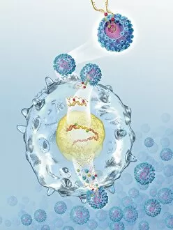
HIV invading white blood cell. Computer artwork showing HIV (human immunodeficiency virus) particles (virions, purple) invading a white blood cell (large, centre) in the human bloodstream

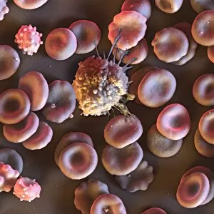
Background imageCytological Collection: Blood cells, SEM C016 / 8026

Blood cells, SEM C016 / 8026
Blood cells. Coloured scanning electron micrograph (SEM) of a lymphocyte white blood cell (centre) and red blood cells (erythrocytes, circular)

Background imageCytological Collection: MDCK culture cells, SEM C016 / 8028

MDCK culture cells, SEM C016 / 8028
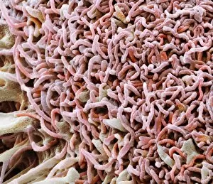
MDCK cells. Coloured scanning electron micrograph (SEM) of cultured MDCK (Madin-Darby canine kidney) epithelial cells on filter paper (fibres)

Background imageCytological Collection: Red blood cells, SEM C016 / 8027

Red blood cells, SEM C016 / 8027
Red blood cells. Coloured scanning electron micrograph (SEM) of red blood cells (erythrocytes). The main function of red blood cells is to distribute oxygen to body tissues

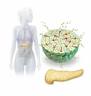
Background imageCytological Collection: Insulin production, artwork

Insulin production, artwork
Insulin production. Computer artwork of the human pancreas (yellow) and an islet of Langerhans (green) producing the hormone insulin (small spheres)

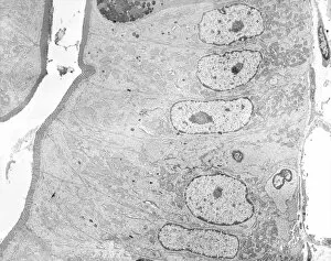
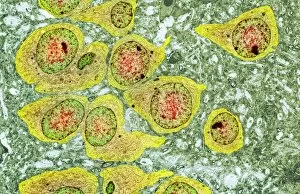
Background imageCytological Collection: Brain nerve cells, TEM C014 / 0356

Brain nerve cells, TEM C014 / 0356
Brain nerve cells. Transmission electron micrograph (TEM) of a section through brain tissue from the cerebral cortex, showing numerous neurons (nerve cells) surrounded by axons and dendrites

Background imageCytological Collection: Brain cell, TEM C014 / 0358

Brain cell, TEM C014 / 0358
Brain cell. Transmission electron micrograph (TEM) of a section through an oligodendrocyte in human brain tissue. Oligodendrocytes occur in both the white

Background imageCytological Collection: Dividing cancer cell, SEM C014 / 0361

Dividing cancer cell, SEM C014 / 0361
Dividing cancer cell. Coloured scanning electron micrograph (SEM) of a colorectal cancer cell undergoing mitosis (nuclear division) and splitting into two daughter cells (left and right)

Background imageCytological Collection: Brain cell, TEM C014 / 0359

Brain cell, TEM C014 / 0359
Brain cell. Transmission electron micrograph (TEM) of a section through an oligodendrocyte in human brain tissue. Oligodendrocytes occur in both the white

Background imageCytological Collection: Dividing brain cancer cells, SEM C014 / 0354

Dividing brain cancer cells, SEM C014 / 0354
Dividing brain cancer cells. Coloured scanning electron micrograph (SEM) of a cancerous astrocyte brain cell that has just undergone cytokinesis (cell division)


For sale as Licensed Images

Choose your image, Select your licence and Download the media

"Cytological Wonders Unveiled: Exploring the Intricacies of Cellular Life" In this captivating journey into the microscopic world, we delve into the realm of cytology, where astonishing discoveries await. From anaesthetic inhibiting an ion channel to mesmerizing light and electron micrographs, each image offers a glimpse into the intricate workings of cells. Firstly, we witness an extraordinary phenomenon as anaesthetic molecules inhibit an ion channel within cells. This interaction sheds light on how these substances affect cellular processes and highlights their potential therapeutic applications. Moving forward, our attention is drawn to HeLa cells captured under a light microscope. The vivid details reveal their complex structures and provide valuable insights for research in various fields such as cancer biology and drug development. Next up is a stunning transmission electron micrograph showcasing the rough endoplasmic reticulum. Its labyrinthine network serves as a hub for protein synthesis and transport within cells, emphasizing its crucial role in maintaining cellular homeostasis. As we shift gears towards artistic representation, dendritic cells take center stage through exquisite artwork. These specialized immune cells play a pivotal role in recognizing foreign invaders and orchestrating immune responses - truly nature's defenders at work. Returning to HeLa cells under another light microscope lens unveils yet another breathtaking display of cellular beauty. Each cell appears like a universe unto itself with intricate organelles working harmoniously to sustain life's delicate balance. The enchantment continues with captivating images capturing mitosis - the process by which one cell divides into two identical daughter cells. Witnessing this dance of chromosomes during cell division provides profound insights into growth, development, and regeneration mechanisms within organisms. Shifting focus from animal to plant life brings us pine pollen grains delicately observed under a light microscope. These tiny particles hold immense significance in plant reproduction dynamics while exhibiting remarkable structural intricacies that aid successful pollination. Further exploring plants' hidden wonders takes us on an enlightening journey through light micrographs of pine and lime tree stems.