
Dentistry Collection (#6)
"Dentistry: A Window into the World of Oral Health" Step into the whimsical world of dentistry, where imagination and innovation collide


For sale as Licensed Images
Choose your image, Select your licence and Download the media
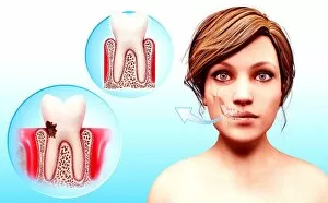
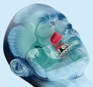
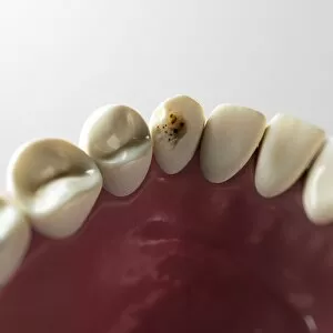
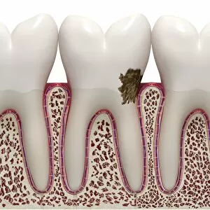
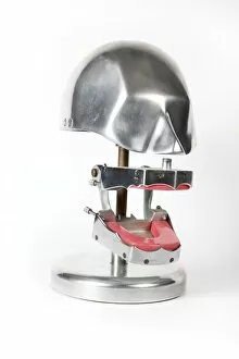
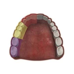
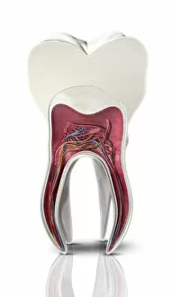
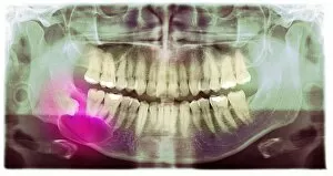
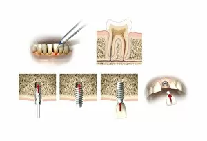
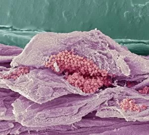
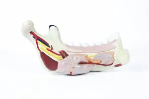
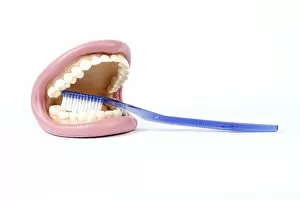
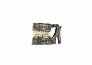
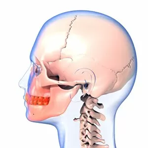
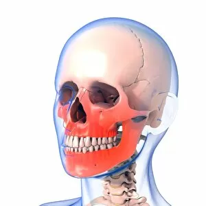
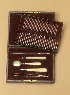
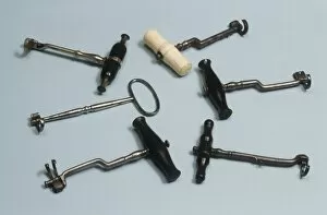
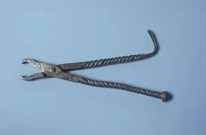
"Dentistry: A Window into the World of Oral Health" Step into the whimsical world of dentistry, where imagination and innovation collide. Bound to Draw by William Heath Robinson, this captivating art piece showcases the intricate machinery behind dental procedures, reminding us that even in complexity lies beauty. A panoramic dental X-ray reveals a hidden universe within our mouths – a mesmerizing landscape of teeth, both adult and childlike. It reminds us that oral health knows no age boundaries; it is an essential aspect of overall well-being. In H. M. Bateman's cartoon "Cause & Effect, " we witness the comical side as a patient's reaction mirrors their dentist's actions. Laughter fills the room as we realize that humor can be found even in seemingly daunting situations. False teeth, meticulously crafted to restore smiles lost along life's journey, offer hope and confidence to those who wear them, and are not just replacements but symbols of resilience and second chances. Witnessing a tooth being filled evokes awe at modern dentistry's ability to repair what was once broken or decayed. The Italian dentist in Naples exemplifies how oral care transcends borders, bringing smiles worldwide. Peering through a dentist's window adorned with countless teeth molds in Zhongdian captures history frozen in time – each mold representing someone whose smile has been transformed by skilled hands. Toy teeth remind us that learning about oral hygiene starts from childhood – instilling habits for lifelong dental health while making education playful and engaging for young minds. Polarised LM image showcasing tooth decay serves as a stark reminder that neglecting oral health can have consequences beyond aesthetics - emphasizing prevention as key to maintaining healthy smiles. The Dentist Chair by Claudius Ash Sons & Co Ltd stands tall as an emblematic symbol of comfort amidst anxiety-filled visits - offering reassurance during routine check-ups or complex treatments alike.