Dentistry Collection (#9)

"Dentistry: A Window into the World of Oral Health" Step into the whimsical world of dentistry, where imagination and innovation collide

Background imageDentistry Collection: An itinerant tooth-puller. Line engraving, Dutch, 18th century

An itinerant tooth-puller. Line engraving, Dutch, 18th century
DENTISTRY, 18th CENTURY. An itinerant tooth-puller. Line engraving, Dutch, 18th century

Background imageDentistry Collection: An itenerant tooth-puller. Line engraving, Dutch, 18th century

An itenerant tooth-puller. Line engraving, Dutch, 18th century
DENTIST, 18TH CENTURY An itenerant tooth-puller. Line engraving, Dutch, 18th century

Background imageDentistry Collection: (1819-1868). American dentist. Dr. Morton successfully performing a tooth extraction under ether

(1819-1868). American dentist. Dr. Morton successfully performing a tooth extraction under ether anesthetic on 30
WILLIAM T.G. MORTON (1819-1868). American dentist. Dr. Morton successfully performing a tooth extraction under ether anesthetic on 30 September 1846, in Boston, Massachuesetts

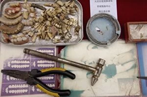
Background imageDentistry Collection: Local dentist in Xizhou market. This small Bai village is about 20 km north of Dali

Local dentist in Xizhou market. This small Bai village is about 20 km north of Dali. CHINA

Background imageDentistry Collection: DENTURES. Line engraving, n. d

DENTURES. Line engraving, n. d
DENTURES. Line engraving, n.d

Background imageDentistry Collection: DENTURES, 1853. Line engraving, 1853

DENTURES, 1853. Line engraving, 1853

Background imageDentistry Collection: Advert for Erasmic tooth paste, 1921

Advert for Erasmic tooth paste, 1921 Date: 1921

Background imageDentistry Collection: FONWEISS TOOTHPASTE, 1887. American advertisement, 1887

FONWEISS TOOTHPASTE, 1887. American advertisement, 1887

Background imageDentistry Collection: Tooth dentine, SEM

Tooth dentine, SEM
Tooth dentine. Coloured scanning electron micrograph (SEM) of dentine from a milk tooth. The bulk of a tooth is formed of dentine, a mineralised connective tissue with a similar composition to bone

Background imageDentistry Collection: Tooth resorption cell, SEM

Tooth resorption cell, SEM
Tooth resorption cell. Coloured scanning electron micrograph (SEM) of an odontoclast, a cell that facilitates the process of resorption of milk teeth roots

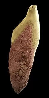
Background imageDentistry Collection: Milk tooth, SEM

Milk tooth, SEM
Milk tooth. Coloured scanning electron micrograph (SEM) of a milk tooth. The root of this tooth has been dissolved, a process known as resorption, leaving just the crown

Background imageDentistry Collection: Molar tooth, SEM

Molar tooth, SEM

Background imageDentistry Collection: Dentin tooth tissue, SEM

Dentin tooth tissue, SEM
Dentin tooth tissue. Coloured scanning electron micrograph (SEM) of dentin tooth tissue. Dentin is mineralised connective tissue that makes up the majority of a tooth

Background imageDentistry Collection: Human canine milk tooth, SEM

Human canine milk tooth, SEM
Milk tooth. Coloured scanning electron micrograph (SEM) of a human canine milk tooth. Milk teeth, also known as deciduous teeth, are the first set of teeth in mammals

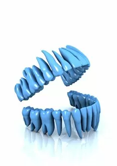
Background imageDentistry Collection: Set of teeth

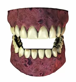
Set of teeth, computer artwork

Background imageDentistry Collection: Tooth decay, SEM

Tooth decay, SEM
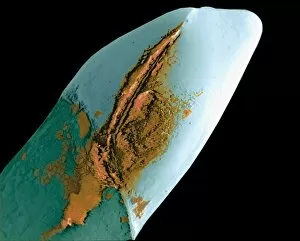
Tooth decay. Coloured scanning electron micrograph (SEM) of dental plaque (brown) on a human incisor. Plaque is a film of bacteria embedded in a glycoprotein matrix

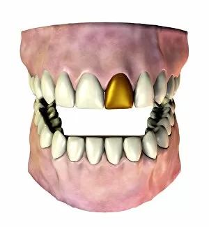
Background imageDentistry Collection: Gold tooth

Gold tooth. Computer artwork of a set of adult teeth, where one of the incisors has been replaced with a gold tooth. Teeth may need to be replaced due to decay or injury

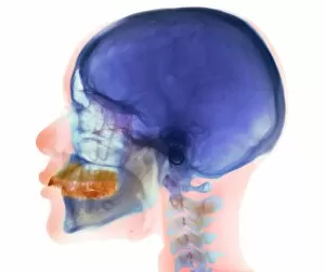
Background imageDentistry Collection: Overbite, X-ray

Overbite, X-ray
Overbite. Coloured X-ray (side view) of the head of a 32 year old woman showing overbite of the upper jaw (yellow) and its teeth

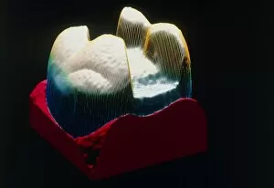
Background imageDentistry Collection: Coloured SEM of a cut dental inlay

Coloured SEM of a cut dental inlay
Dental inlay. (Image 3 of 3). Coloured scanning electron micrograph (SEM) of a dental inlay (blue) on a mount (brown). The inlay has just come from the miniature milling machine in which it was cut

Background imageDentistry Collection: Dental pin, SEM

Dental pin, SEM
Dental pin. Coloured scanning electron micrograph (SEM) of a dental pin. This type of pin is used to keep false teeth in place

Background imageDentistry Collection: Used and unused toothpicks, SEM

Used and unused toothpicks, SEM
Used and unused toothpicks. Coloured scanning electron micrograph (SEM) of an unused (left) and a used (right) toothpicks. The used toothpick has a blunt end and is covered in dental plaque

Background imageDentistry Collection: X-ray of teeth

X-ray of teeth
False-colour dental x-ray of teeth from the right hand side of a 45 year old male mouth. The red areas are fillings, while the green area in the tooth at bottom left is a patch of caries

Background imageDentistry Collection: Used toothbrush bristle, SEM

Used toothbrush bristle, SEM
Used toothbrush bristle. Coloured scanning electron micrograph (SEM) of dental plaque on a bristle from a used toothbrush. Plaque consists of a film of bacteria embedded in a glycoprotein matrix

Background imageDentistry Collection: Extracted molar

Extracted molar
Macrophotograph of an extracted molar (male, upper, number 7), showing the build up of tartar, formed from salts in the saliva, around the gum line (brown line). Magnification: X1.2 (at 35mm size)

Background imageDentistry Collection: Root-canal treatment, dental X-ray

Root-canal treatment, dental X-ray
Dental root-canal treatment. Coloured pan-oral X- ray of the mouth of a 17 year old woman showing a number of fillings (green) in teeth of the upper jaw, including root-canal treatment

Background imageDentistry Collection: Coloured X-ray of impacted wisdom tooth and brace

Coloured X-ray of impacted wisdom tooth and brace
Impacted wisdom tooth. Coloured X-ray of the mouth showing an impacted wisdom tooth (highlighted yellow/magenta, centre left)

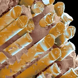
Background imageDentistry Collection: Coloured SEM of a glass fibre dental ceramic

Coloured SEM of a glass fibre dental ceramic
Glass fibre dental ceramic. Coloured scanning electron micrograph (SEM) of a dental ceramic reinforced with glass fibres. This ceramic is used to make artificial teeth such as those used in dental

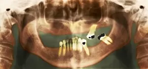
Background imageDentistry Collection: Loss of teeth, X-ray

Loss of teeth, X-ray
Loss of teeth. Coloured pan-oral X-ray of the mouth of an elderly man showing his few remaining teeth. Some of the teeth (for instance at centre right) have fillings (black) in them

Background imageDentistry Collection: Polarised LM of a tooth with a dental crown

Polarised LM of a tooth with a dental crown
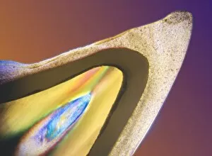
Dental crown. Polarised light micrograph (PLM) of a longitudinal section through a tooth (yellow & blue) with a dental crown (grainy area)

Background imageDentistry Collection: Gum disease

Gum disease. Computer artwork of diseased gums and teeth. Gum disease causes inflammation and bleeding of the gums. It is the most common cause of tooth loss

Background imageDentistry Collection: Coloured SEM of a dental drill drilling into tooth

Coloured SEM of a dental drill drilling into tooth
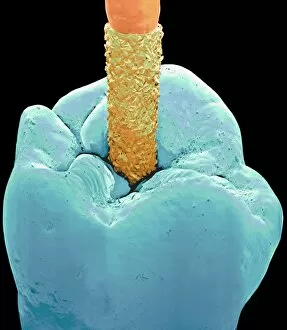
Dental drill on tooth. Coloured scanning electron micrograph (SEM) of a diamond-tipped dental drill (orange) drilling a hole into a tooth (blue)

Background imageDentistry Collection: Coloured SEM of a tooth with a dental crown

Coloured SEM of a tooth with a dental crown
Dental crown. Coloured scanning electron micrograph (SEM) of a tooth (green) with a dental crown (light blue). The tooth and ceramic crown are coated in debris (orange)

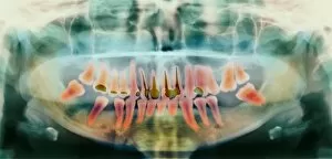
Background imageDentistry Collection: Dental X-ray

Dental X-ray
MODEL RELEASED. Dental X-ray. Dentist looking at an X-ray of a patients teeth. This is a panoramic wraparound X-ray that shows all the teeth of both the upper and lower jaw

Background imageDentistry Collection: Coloured SEM of the join between a tooth and crown

Coloured SEM of the join between a tooth and crown
Dental crown. Coloured scanning electron microgr- aph (SEM) of the join between a tooth (green) and a dental crown (light blue)

Background imageDentistry Collection: Artwork of tooth showing periodontal disease

Artwork of tooth showing periodontal disease
Periodontal disease. Artwork of a section through a human tooth with periodontal disease of its surrounding gum and bone. The tooths white enamel covers the dentine (yellow) and pulp (purple)

Background imageDentistry Collection: Dental drill, SEM

Dental drill, SEM
Dental drill. Coloured scanning electron micrograph (SEM) of the tip of a dental drill bit. Magnification: x28 when printed at 10 centimetres wide

Background imageDentistry Collection: Decay on molar tooth, SEM

Decay on molar tooth, SEM
Tooth decay. Scanning electron micrograph (SEM) of the surface of a decaying molar tooth. The surface enamel (white) has begun to break down (for example, at lower right)

Background imageDentistry Collection: Dental inlay being cut in a milling machine

Dental inlay being cut in a milling machine

Background imageDentistry Collection: Extracted molar showing 3 amalgam fillings

Extracted molar showing 3 amalgam fillings
Macrophotograph of an extracted molar (male, upper, number 7), a top view showing three 20 year old amalgam fillings. The filling is a powdered alloy consisting of silver, tin

Background imageDentistry Collection: Computer graphic of a tooth ready for dental inlay

Computer graphic of a tooth ready for dental inlay
Dental inlay preparation. (Image 1 of 3). Computer graphic of a tooth with a cavity cut into it in preparation for a dental inlay

Background imageDentistry Collection: Coloured SEM of the root of a dental implant

Coloured SEM of the root of a dental implant

Background imageDentistry Collection: Dental plaque

Dental plaque
Tooth plaque. Dental plaque (yellow) on a patients teeth. Plaque is the main cause of tooth decay. It is a sticky coating that consists of saliva, bacteria and particles of food

Background imageDentistry Collection: Complete loss of teeth, X-ray

Complete loss of teeth, X-ray
Loss of teeth. Coloured X-ray (side view) of the head of a 49 year old woman showing complete loss of teeth in both upper and lower jaws

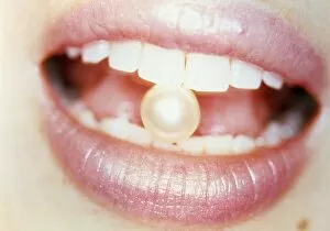
Background imageDentistry Collection: Pearl between teeth

Pearl between teeth. Conceptual image of a pearl held between a womans teeth. This could represent teeth that are clean, healthy, white, polished or " pearly"

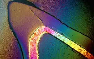
Background imageDentistry Collection: Polarised LM of a cracked ceramic dental crown

Polarised LM of a cracked ceramic dental crown
Dental crown. Polarised light micrograph (PLM) of a longitudinal section through a cracked dental crown made of metal (multicoloured) and ceramic (above metal)

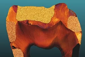
Background imageDentistry Collection: Polarised LM of a tooth with various fillings

Polarised LM of a tooth with various fillings
Tooth fillings. Polarised light micrograph (PLM) of a longitudinal section through a tooth with various fillings. The enamel (orange) has been replaced in areas of decay (dental caries)

Background imageDentistry Collection: Trapped food in dental braces

Trapped food in dental braces
MODEL RELEASED. Trapped food in dental braces, which are fixed to the teeth of a teenage girl. Fixed braces consist of metal brackets cemented to each tooth

Background imageDentistry Collection: Toothpaste, SEM

Toothpaste, SEM
Toothpaste, coloured scanning electron micrograph (SEM)


For sale as Licensed Images

Choose your image, Select your licence and Download the media

"Dentistry: A Window into the World of Oral Health" Step into the whimsical world of dentistry, where imagination and innovation collide. Bound to Draw by William Heath Robinson, this captivating art piece showcases the intricate machinery behind dental procedures, reminding us that even in complexity lies beauty. A panoramic dental X-ray reveals a hidden universe within our mouths – a mesmerizing landscape of teeth, both adult and childlike. It reminds us that oral health knows no age boundaries; it is an essential aspect of overall well-being. In H. M. Bateman's cartoon "Cause & Effect, " we witness the comical side as a patient's reaction mirrors their dentist's actions. Laughter fills the room as we realize that humor can be found even in seemingly daunting situations. False teeth, meticulously crafted to restore smiles lost along life's journey, offer hope and confidence to those who wear them, and are not just replacements but symbols of resilience and second chances. Witnessing a tooth being filled evokes awe at modern dentistry's ability to repair what was once broken or decayed. The Italian dentist in Naples exemplifies how oral care transcends borders, bringing smiles worldwide. Peering through a dentist's window adorned with countless teeth molds in Zhongdian captures history frozen in time – each mold representing someone whose smile has been transformed by skilled hands. Toy teeth remind us that learning about oral hygiene starts from childhood – instilling habits for lifelong dental health while making education playful and engaging for young minds. Polarised LM image showcasing tooth decay serves as a stark reminder that neglecting oral health can have consequences beyond aesthetics - emphasizing prevention as key to maintaining healthy smiles. The Dentist Chair by Claudius Ash Sons & Co Ltd stands tall as an emblematic symbol of comfort amidst anxiety-filled visits - offering reassurance during routine check-ups or complex treatments alike.