
Disease Collection (page 100)
"Disease: A Historical Battle Against the Unseen Foe" In 1845, as the HMS Erebus and HMS Terror set sail on their ill-fated Arctic expedition


For sale as Licensed Images
Choose your image, Select your licence and Download the media
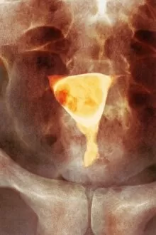
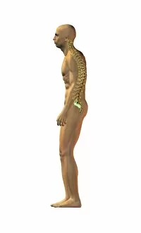
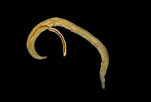
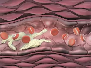
"Disease: A Historical Battle Against the Unseen Foe" In 1845, as the HMS Erebus and HMS Terror set sail on their ill-fated Arctic expedition, little did they know that disease would become their deadliest adversary. The Shotley Bridge General Hospital in County Durham witnessed countless patients afflicted by mysterious ailments brought back from distant lands. Captain Francis Crozier of HMS Terror valiantly led his crew through treacherous waters, but even his courage could not shield them from the invisible enemy lurking within. As depicted in a Punch cartoon, renowned scientist Faraday tried to offer his expertise to Father Thames, symbolizing society's desperate attempts to combat diseases. The battle against disease takes place at microscopic levels too. T lymphocytes engaging cancer cells under an electron microscope (SEM C001 / 1679) exemplify our ongoing fight against this relentless foe. Awareness campaigns remind us that "Coughs and Sneezes Spread Diseases, " urging us to take precautions for public health. Medical advancements have revolutionized treatments over time. Pneumothorax treatment using X-rays has saved countless lives by effectively collapsing lung cavities caused by diseases like tuberculosis. However, these triumphs cannot overshadow the tragedies faced by Captain Sir John Franklin and his crew who succumbed to disease during their Arctic exploration. Diseases have often been personified throughout history – King Cholera presiding over a court reminds us of the devastating cholera outbreaks that plagued communities worldwide. Medical illustrations depicting appendicitis or bunions captured the urgency with which doctors sought solutions for these painful conditions. Yet perhaps one of the most feared diseases is Alzheimer's, which silently erodes memories and identities. Computer artwork visualizing an Alzheimer's brain serves as a stark reminder of its impact on individuals and families alike. As we navigate through centuries battling various diseases, it becomes evident that our fight is far from over.