Disease Collection (#93)

"Disease: A Historical Battle Against the Unseen Foe" In 1845, as the HMS Erebus and HMS Terror set sail on their ill-fated Arctic expedition

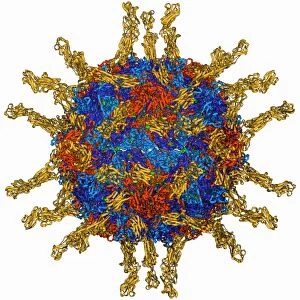
Background imageDisease Collection: Semliki forest virus capsid F006 / 9297

Semliki forest virus capsid F006 / 9297
Semliki forest virus capsid, molecular model. This virus, named for the forest in Uganda where it was identified, is spread by the bite of mosquitoes. It can infect both humans and animals

Background imageDisease Collection: Human poliovirus, molecular model F006 / 9289

Human poliovirus, molecular model F006 / 9289
Human poliovirus particle. Molecular model of the capsid of the human poliovirus. The capsid is a protein coat that encloses the viruss genetic information (genome), stored as RNA (ribonucleic acid)

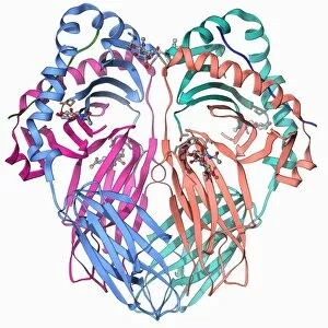
Background imageDisease Collection: MHC protein complexed with flu virus F006 / 9294

MHC protein complexed with flu virus F006 / 9294
MHC protein complexed with flu virus. Molecular model showing human class II MHC (major histocompatibility complex) protein HLA-DR1 complexed with an influenza (flu) virus peptide

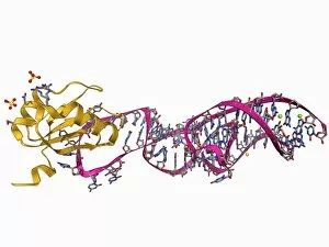
Background imageDisease Collection: Hepatitis D virus ribozyme complex F006 / 9295

Hepatitis D virus ribozyme complex F006 / 9295
Hepatitis D virus ribozyme complex. Molecular model showing an RNA (ribonucleic acid) strand from an Hepatitis delta (Hepatitis D) virus genomic ribozyme, complexed with a ribonucleoprotein

Background imageDisease Collection: Human interferon beta molecule F006 / 9241

Human interferon beta molecule F006 / 9241
Human interferon beta, molecular model. Interferons are proteins produced by white blood cells as part of the immune response to invading pathogens, especially viruses

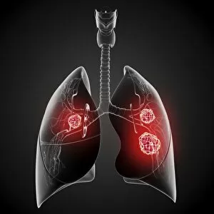
Background imageDisease Collection: Female lung cancer, artwork

Female lung cancer, artwork
Female lung cancer, computer artwork

Background imageDisease Collection: Amyloid precursor protein molecule F006 / 9224

Amyloid precursor protein molecule F006 / 9224
Amyloid precursor protein. Molecular model showing the structure of the protease inhibitor domain of an amyloid precursor protein (APP)

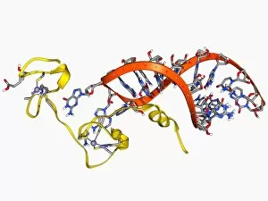
Background imageDisease Collection: HIV nucleocapsid protein molecule F006 / 9219

HIV nucleocapsid protein molecule F006 / 9219
HIV nucleocapsid protein. Molecular model of the nucleocapsid protein (yellow) from HIV-1 (human immunodeficiency virus-type 1) complexed with the Psi RNA (ribonucleic acid) packaging element (orange)

Background imageDisease Collection: Lung cancer, artwork

Lung cancer, artwork
Lung cancer, computer artwork

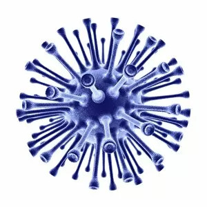
Background imageDisease Collection: H1N1 flu virus particle, artwork F006 / 9186

H1N1 flu virus particle, artwork F006 / 9186
H1N1 flu virus particle. Computer artwork of an H1N1 influenza A (flu) virus particle (virion). In the particles lipid envelope (purple) are two types of protein spike

Background imageDisease Collection: Coronary stent, X-ray F006 / 9141

Coronary stent, X-ray F006 / 9141
Coronary stent. Coloured angiogram (blood vessel X-ray) of the coronary arteries of a 52 year old patient. The artery at right has had a stent placed in it to treat a blockage

Background imageDisease Collection: Knee disease, CT scan F006 / 9140

Knee disease, CT scan F006 / 9140
Knee disease. Coloured axial computer tomography (CT) scan of the right knee of an 18 year old patient with osteochondritis dissecans (OCD) of the knee

Background imageDisease Collection: Knee disease, X-ray F006 / 9138

Knee disease, X-ray F006 / 9138
Knee disease. Coloured X-ray of the right knee of an 18 year old patient with osteochondritis dissecans (OCD) of the knee

Background imageDisease Collection: Murine minute virus capsid

Murine minute virus capsid, molecular model. This parvovirus infects mice, its only known natural host. It is highly infectious, transmitted through the nose and mouth

Background imageDisease Collection: Scoliosis of the spine, X-ray F006 / 9134

Scoliosis of the spine, X-ray F006 / 9134
Scoliosis of the spine. Coloured X-ray of the chest of a 31 year old patient with scoliosis (sideways curvature) of the spine

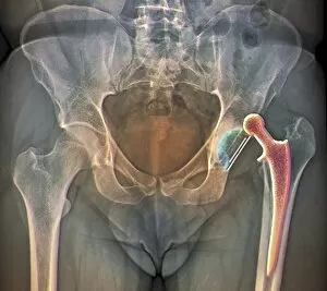
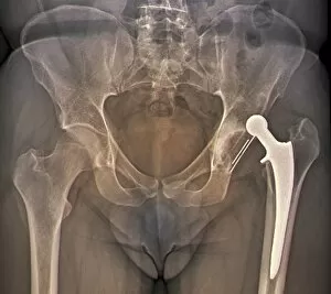
Background imageDisease Collection: Dislocated hip replacement, X-ray F006 / 9132

Dislocated hip replacement, X-ray F006 / 9132
Dislocated hip replacement. Coloured frontal X-ray of the pelvis of a 35 year old patient with an artificial left hip (right) that has become dislocated

Background imageDisease Collection: Scoliosis of the spine, X-ray F006 / 9133

Scoliosis of the spine, X-ray F006 / 9133
Scoliosis of the spine. Coloured X-ray of the chest of a 31 year old patient with scoliosis (sideways curvature) of the spine

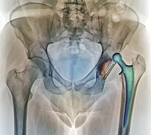
Background imageDisease Collection: Dislocated hip replacement, X-ray F006 / 9131

Dislocated hip replacement, X-ray F006 / 9131
Dislocated hip replacement. Coloured frontal X-ray of the pelvis of a 35 year old patient with an artificial left hip (right) that has become dislocated

Background imageDisease Collection: Dislocated hip replacement, X-ray F006 / 9130

Dislocated hip replacement, X-ray F006 / 9130
Dislocated hip replacement. Coloured frontal X-ray of the pelvis of a 35 year old patient with an artificial left hip (right) that has become dislocated

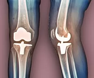
Background imageDisease Collection: Total knee replacement, X-rays F006 / 9123

Total knee replacement, X-rays F006 / 9123
Total knee replacement. Coloured frontal (left) and profile (right) X-rays of the right knee of a 69 year old patient after total knee replacement surgery

Background imageDisease Collection: Total knee replacement, X-rays F006 / 9122

Total knee replacement, X-rays F006 / 9122
Total knee replacement. Coloured frontal (left) and profile (right) X-rays of the right knee of a 69 year old patient after total knee replacement surgery

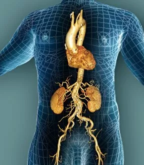
Background imageDisease Collection: Aorta, 3D CT scan F006 / 9120

Aorta, 3D CT scan F006 / 9120
Aorta. Coloured 3D computed tomography (CT) angiogram of the aorta of a 54 year old patient. The aorta is the bodys main artery

Background imageDisease Collection: Aorta, 3D CT scan F006 / 9119

Aorta, 3D CT scan F006 / 9119
Aorta. Coloured 3D computed tomography (CT) angiogram of the aorta of a 54 year old patient. The aorta is the bodys main artery

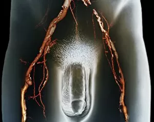
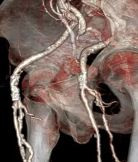
Background imageDisease Collection: Narrowed arteries, 3D CT scan F006 / 9113

Narrowed arteries, 3D CT scan F006 / 9113
Narrowed arteries. Coloured 3D computer tomography (CT) angiogram of the pelvis of a 66 year old male patient with narrowing (stenosis) of the iliac and femoral arteries

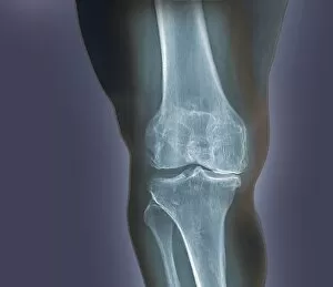
Background imageDisease Collection: Arthritis of the knee, X-ray F006 / 9104

Arthritis of the knee, X-ray F006 / 9104
Arthritis of the knee. Coloured X-ray of the knee of an obese 48 year old patient showing degenerative arthritis caused by the extreme stress on the knee from being overweight

Background imageDisease Collection: Arthritis of the knee, X-ray F006 / 9103

Arthritis of the knee, X-ray F006 / 9103
Arthritis of the knee. Coloured X-ray of the knee of an obese 48 year old patient showing degenerative arthritis caused by the extreme stress on the knee from being overweight

Background imageDisease Collection: Septic arthritis, X-ray C017 / 7582

Septic arthritis, X-ray C017 / 7582
Septic arthritis. X-ray of a side view of the shoulder joint of a 24 year old male that has been damaged due to septic arthritis

Background imageDisease Collection: Atherosclerosis, artwork

Atherosclerosis, artwork
Atherosclerosis. Computer artwork of a narrowed artery, due to a cholesterol plaque

Background imageDisease Collection: SV40 virus capsid, molecular model C018 / 7904

SV40 virus capsid, molecular model C018 / 7904
SV40 virus capsid, molecular model. Simian virus 40 (SV40) is found in monkeys such as Rhesus monkeys and macaques. Potentially tumour-causing, it is used in laboratory research and in vaccines

Background imageDisease Collection: SV40 virus capsid, molecular model C018 / 7903

SV40 virus capsid, molecular model C018 / 7903
SV40 virus capsid, molecular model. Simian virus 40 (SV40) is found in monkeys such as Rhesus monkeys and macaques. Potentially tumour-causing, it is used in laboratory research and in vaccines

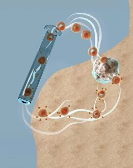
Background imageDisease Collection: Immunotherapy, artwork

Immunotherapy, artwork
Immunotherapy. Computer artwork of immunotherapy being used to destroy a tumour (right). Immunotherapy uses the bodys immune system to fight a disease

Background imageDisease Collection: Thrombophlebitis in the brain, MRI scans

Thrombophlebitis in the brain, MRI scans
Thrombophlebitis in the brain. Coloured magnetic resonance imaging (MRI) angiograms comparing the blood vessels (white) of the brain and neck in a healthy 43-year-old (left)

Background imageDisease Collection: Migraine, conceptual image

Migraine, conceptual image

Background imageDisease Collection: Thrombophlebitis in the brain, 3D CT scan

Thrombophlebitis in the brain, 3D CT scan
Thrombophlebitis in the brain. Coloured 3D computed tomography (CT) angiogram of the blood vessels (blue) in the brain of a 48-year-old with thrombophlebitis of the superior sagittal sinus

Background imageDisease Collection: Pouchitis, endoscope view C015 / 5074

Pouchitis, endoscope view C015 / 5074
Pouchitis, endoscope view. This is the inflammation of an artificial rectum, known as an ileal pouch, formed in patients that have had their large intestine removed

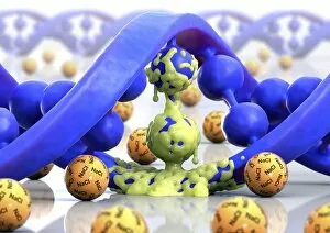
Background imageDisease Collection: Cystic fibrosis, conceptual artwork C015 / 5022

Cystic fibrosis, conceptual artwork C015 / 5022
Cystic fibrosis, conceptual computer artwork. Molecule of DNA (deoxyribonucleic acid, blue) partly covered in mucus (yellow) surrounded by salt (NaCl) molecules

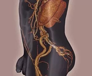
Background imageDisease Collection: Atherosclerosis, 3D CT scan

Atherosclerosis, 3D CT scan
Atherosclerosis. 3D computed tomography (CT) angiogram of the main blood vessels of the lower body a 58-year-old man, showing numerous atheromatous plaques (light, centre) on the vessel walls

Background imageDisease Collection: Human head, MRI and 3D CT scans

Human head, MRI and 3D CT scans
Stroke. Coloured composite image of a magnetic resonance imaging (MRI) scan of the brain and 2D and 3D computed tomography (CT)

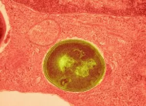
Background imageDisease Collection: Staphylococcus aureus bacteria, TEM C016 / 9458

Staphylococcus aureus bacteria, TEM C016 / 9458
Staphylococcus aureus bacteria. Coloured transmission electron micrograph (TEM) of a Staphylococcus aureus bacterium (round) in skin tissue. S

Background imageDisease Collection: Flu virus, conceptual artwork C016 / 7530

Flu virus, conceptual artwork C016 / 7530
Flu virus, conceptual computer artwork

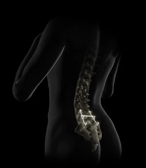
Background imageDisease Collection: Lumbar spine fusion, artwork C016 / 6993

Lumbar spine fusion, artwork C016 / 6993
Lumbar spine fusion. Artwork of pins and screws inserted into a patients lumbar spine in a case of spinal fusion. The pins hold together the patients vertebrae

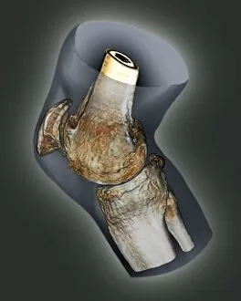
Background imageDisease Collection: Arthritis of the knee, 3D CT scan C016 / 6510

Arthritis of the knee, 3D CT scan C016 / 6510
Arthritis of the knee. Coloured 3D computed tomography (CT) scan of the right knee of a 65 year old patient with severe osteoarthritis

Background imageDisease Collection: Atherosclerosis, 3D CT scan C016 / 6537

Atherosclerosis, 3D CT scan C016 / 6537
Atherosclerosis. Coloured 3D computed tomography (CT) angiogram of the hip of a 67 year old patient with hypertension (high blood pressure) and hypercholesterolemia (high blood cholesterol)

Background imageDisease Collection: Atherosclerosis, 3D CT scan C016 / 6540

Atherosclerosis, 3D CT scan C016 / 6540
Atherosclerosis. Coloured 3D computed tomography (CT) angiogram of the abdomen of a 73 year old patient. The aorta (cream, down centre), the bodys main artery, has calcified fatty plaques (atheroma)

Background imageDisease Collection: Atherosclerosis, 3D CT scan C016 / 6499

Atherosclerosis, 3D CT scan C016 / 6499
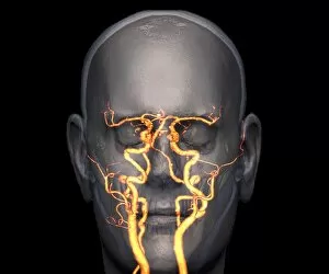
Atherosclerosis. Coloured 3D computed tomography (CT) angiogram of the head of a 60 year old patient with hypertension (high blood pressure) and hypercholesterolemia (high blood cholesterol)

Background imageDisease Collection: Neck arthritis treatment, angiogram C016 / 6440

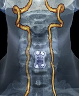
Neck arthritis treatment, angiogram C016 / 6440
Neck arthritis treatment. Coloured X-ray angiogram of the neck of a 48 year old patient with severe osteoarthritis of the neck

Background imageDisease Collection: Arthritis of the knee, 3D CT scan C016 / 6511

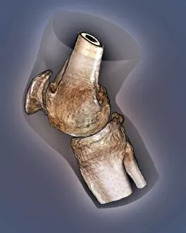
Arthritis of the knee, 3D CT scan C016 / 6511
Arthritis of the knee. Coloured 3D computed tomography (CT) scan of the right knee of a 65 year old patient with severe osteoarthritis

Background imageDisease Collection: Neck arthritis treatment, angiogram C016 / 6443

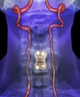
Neck arthritis treatment, angiogram C016 / 6443
Neck arthritis treatment. Coloured X-ray angiogram of the neck of a 48 year old patient with severe osteoarthritis of the neck


For sale as Licensed Images

Choose your image, Select your licence and Download the media

"Disease: A Historical Battle Against the Unseen Foe" In 1845, as the HMS Erebus and HMS Terror set sail on their ill-fated Arctic expedition, little did they know that disease would become their deadliest adversary. The Shotley Bridge General Hospital in County Durham witnessed countless patients afflicted by mysterious ailments brought back from distant lands. Captain Francis Crozier of HMS Terror valiantly led his crew through treacherous waters, but even his courage could not shield them from the invisible enemy lurking within. As depicted in a Punch cartoon, renowned scientist Faraday tried to offer his expertise to Father Thames, symbolizing society's desperate attempts to combat diseases. The battle against disease takes place at microscopic levels too. T lymphocytes engaging cancer cells under an electron microscope (SEM C001 / 1679) exemplify our ongoing fight against this relentless foe. Awareness campaigns remind us that "Coughs and Sneezes Spread Diseases, " urging us to take precautions for public health. Medical advancements have revolutionized treatments over time. Pneumothorax treatment using X-rays has saved countless lives by effectively collapsing lung cavities caused by diseases like tuberculosis. However, these triumphs cannot overshadow the tragedies faced by Captain Sir John Franklin and his crew who succumbed to disease during their Arctic exploration. Diseases have often been personified throughout history – King Cholera presiding over a court reminds us of the devastating cholera outbreaks that plagued communities worldwide. Medical illustrations depicting appendicitis or bunions captured the urgency with which doctors sought solutions for these painful conditions. Yet perhaps one of the most feared diseases is Alzheimer's, which silently erodes memories and identities. Computer artwork visualizing an Alzheimer's brain serves as a stark reminder of its impact on individuals and families alike. As we navigate through centuries battling various diseases, it becomes evident that our fight is far from over.