
Dissected Collection (#3)
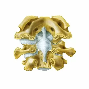
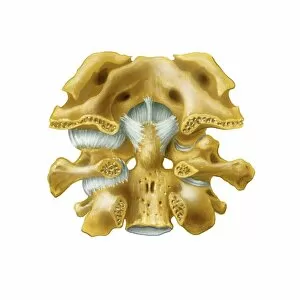
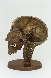
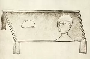
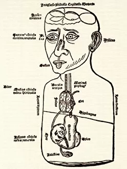
"Unveiling the Intricacies of Life: A Journey through Dissected Hints" Step into the world of Leonardo da Vinci's skull anatomy


For sale as Licensed Images
Choose your image, Select your licence and Download the media
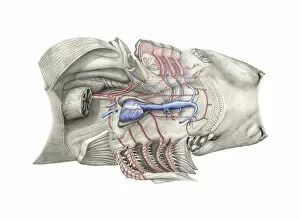
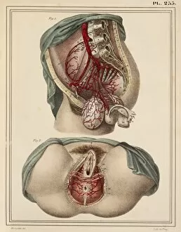
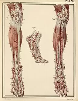
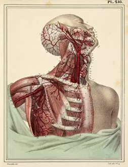
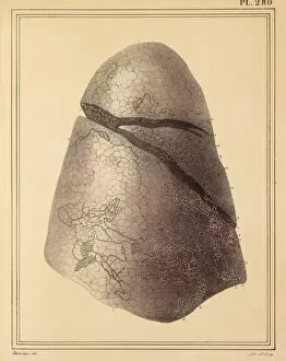
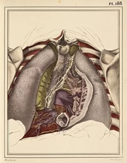
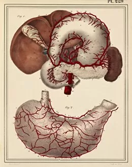
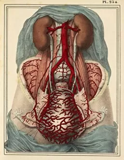
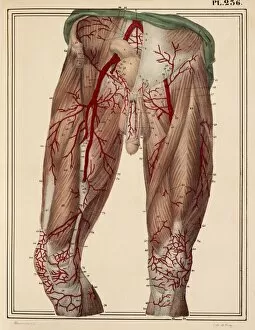
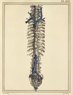
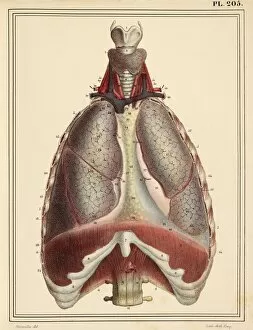
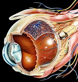
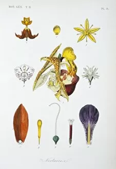
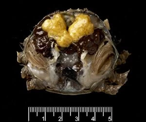
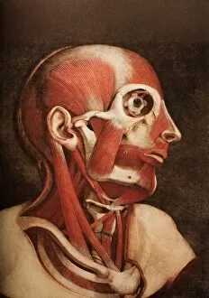
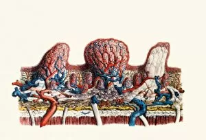
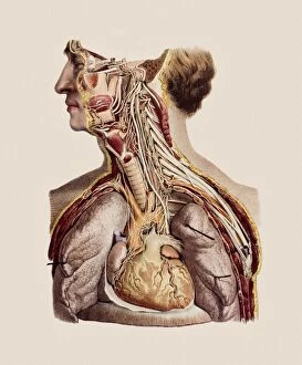
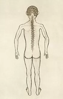
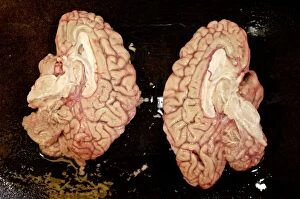
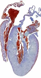
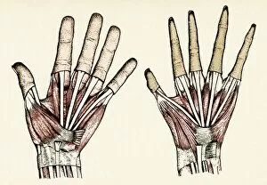
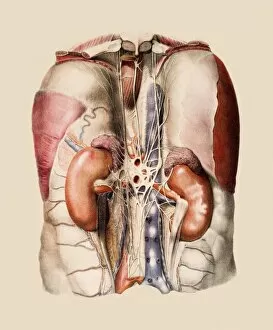
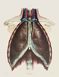
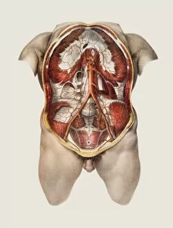
"Unveiling the Intricacies of Life: A Journey through Dissected Hints" Step into the world of Leonardo da Vinci's skull anatomy, where every curve and crevice holds secrets to our existence. Marvel at the intricate network of blood vessels that nourish our brain, as depicted in his mesmerizing artwork. The heart, a symbol of life itself, beats tirelessly within us. Explore its chambers and valves, understanding its vital role in sustaining our being. Witness the delicate dance between arteries and veins that carry life-giving oxygen throughout our bodies. Delve into history with an 1825 artwork showcasing male groin arteries - a testament to early anatomical studies. Discover how these hidden pathways contribute to our overall health and well-being. Venture further into the realm of dissection as we unravel the complex relationship between the heart and lungs. Witness their interdependence, understanding how they work harmoniously to ensure proper oxygenation for survival. Behold an 1825 artwork depicting head and chest arteries; a visual representation of scientific exploration from centuries past. Marvel at how these intricate networks sustain life by delivering essential nutrients throughout our bodies. Explore beyond mere surface appearances as we confront liver cirrhosis – a stark reminder of what can happen when this vital organ is compromised. Gain insight into medical conditions that challenge us while appreciating the resilience of human physiology. Witness scientists meticulously working with ragworm specimens – emblematic of tireless dedication towards unlocking nature's mysteries. Celebrate their unwavering commitment to advancing knowledge for generations to come. Embark on a journey through facial nerves, discovering their role in conveying emotions across countless expressions etched upon our faces. Appreciate how these invisible threads connect mind and body in profound ways. Transport yourself back in time to witness Eustachian tube syringing practices from the 18th century – an intriguing glimpse into historical medical techniques aimed at restoring balance within fragile auditory systems.