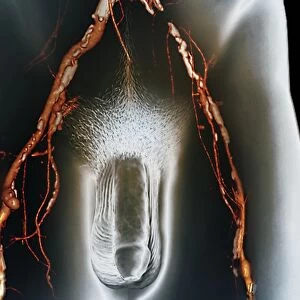
Childs arteries, 1825 artwork

![]()
Wall Art and Photo Gifts from Science Photo Library
Childs arteries, 1825 artwork
Childs arteries. Posterior view of a whole-body dissection showing the arteries (red) of a childs body, with layers of skin and muscles removed to varying depths. Bones are shown at right, including the scapula, pelvis and femur, with more muscles and ligaments present at left. This anatomical artwork is plate 244 from volume 4 of Manuel d anatomie descriptive du corps humain (1825). This 5-volume anatomy atlas was produced by French physician and surgeon Jules Germain Cloquet (1790-1883). The illustrations were by Haincelin. Volume 4 illustrated the anatomy of the circulatory and respiratory systems
Science Photo Library features Science and Medical images including photos and illustrations
Media ID 9222655
© SCIENCE PHOTO LIBRARY
1825 Anatomical Artwork Anatomical Illustration Anatomy Atlas Arms Arterial System Arteries Back Behind Blood Vessels Bones Child Deep Dissected Dissection French Haincelin Jules Germain Cloquet Legs Limbs Muscles Oxygenated Blood Posterior Superficial Vascular Volume 4 Volume Iv Whole Body Artery Blood Vessel Circulatory System
EDITORS COMMENTS
This print showcases a remarkable artwork from 1825 titled "Child's Arteries". The posterior view of a whole-body dissection reveals the intricate network of arteries (highlighted in red) within a child's body. Layers of skin and muscles have been meticulously removed to varying depths, exposing the underlying structures. On the right side, we can observe bones such as the scapula, pelvis, and femur, while more muscles and ligaments are visible on the left. Plate 244 from volume 4 of Manuel d'anatomie descriptive du corps humain is responsible for this stunning anatomical illustration. Created by renowned French physician and surgeon Jules Germain Cloquet with illustrations by Haincelin, this five-volume anatomy atlas delves into the circulatory and respiratory systems' intricacies. The historical significance of this artwork cannot be overstated. It provides us with valuable insights into human anatomy during the early 19th century. By studying these detailed depictions, medical professionals gain a deeper understanding of how blood vessels function within our bodies. This thought-provoking image transports us back in time to witness an era when scientific knowledge was rapidly expanding. It serves as a testament to humanity's relentless pursuit of understanding our own physical selves. Whether you're fascinated by medical history or simply appreciate art that combines beauty with scientific precision, this print is sure to captivate your imagination.
MADE IN THE USA
Safe Shipping with 30 Day Money Back Guarantee
FREE PERSONALISATION*
We are proud to offer a range of customisation features including Personalised Captions, Color Filters and Picture Zoom Tools
SECURE PAYMENTS
We happily accept a wide range of payment options so you can pay for the things you need in the way that is most convenient for you
* Options may vary by product and licensing agreement. Zoomed Pictures can be adjusted in the Cart.