
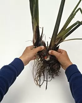
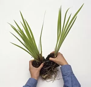
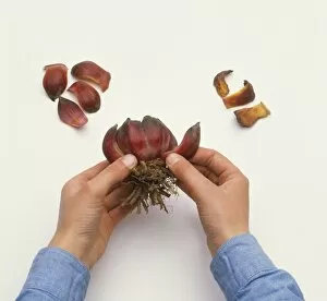
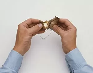
Dividing Collection (page 5)

"Exploring the Intricate Process of Dividing: From Horses to Microorganisms" In a tranquil stable

Background imageDividing Collection: Splitting root mass of Acorus calamus (Sweet flag), using hands, close-up

Splitting root mass of Acorus calamus (Sweet flag), using hands, close-up

Background imageDividing Collection: Hands dividing Iris pseudacorus plant, close-up

Hands dividing Iris pseudacorus plant, close-up

Background imageDividing Collection: Placing daffodil bulb on its cut end, then cutting downwards through basal plate with a sharp knife

Placing daffodil bulb on its cut end, then cutting downwards through basal plate with a sharp knife (propagating by twin-scaling)

Background imageDividing Collection: Dividing pieces of daffodil bulb with scalpel (propagating by twin-scaling)

Dividing pieces of daffodil bulb with scalpel (propagating by twin-scaling)

Background imageDividing Collection: Hands peeling off scales from a lily bulb (propagating by scaling), close-up

Hands peeling off scales from a lily bulb (propagating by scaling), close-up

Background imageDividing Collection: Separating pairs or clumps of daffodil bulbs with large offsets into individual bulbs

Separating pairs or clumps of daffodil bulbs with large offsets into individual bulbs, pulling them apart with fingers, close-up

Background imageDividing Collection: Hands dividing clumps of Nerine bulbs, close-up

Hands dividing clumps of Nerine bulbs, close-up

Background imageDividing Collection: Dividing up clump of Primula denticulata shoots (Drumstick primrose), close-up

Dividing up clump of Primula denticulata shoots (Drumstick primrose), close-up

Background imageDividing Collection: Cutting lengths of root of Primula denticulata (Drumstick primrose), close-up

Cutting lengths of root of Primula denticulata (Drumstick primrose), close-up

Background imageDividing Collection: Propagating by division, loosening the root mass of Gentiana acaulis (Gentian)

Propagating by division, loosening the root mass of Gentiana acaulis (Gentian) and dividing clump into pieces, using hand forks back-to-back, close-up

Background imageDividing Collection: Hands separating rooted Chrysanthemum cuttings, close-up

Hands separating rooted Chrysanthemum cuttings, close-up

Background imageDividing Collection: Cutting off roots from Acanthus, close to crown, to create cuttings, close-up

Cutting off roots from Acanthus, close to crown, to create cuttings, close-up

Background imageDividing Collection: Dividing peonies, dusting divided sections of crown with fungicide

Dividing peonies, dusting divided sections of crown with fungicide, to discourage infection and rot, close-up

Background imageDividing Collection: Using fingers to divide Bergenia into smaller clumps

Using fingers to divide Bergenia into smaller clumps

Background imageDividing Collection: Cynara cardunculus (Artichoke), dividing plant with spade, close-up

Cynara cardunculus (Artichoke), dividing plant with spade, close-up

Background imageDividing Collection: Laurus nobilis (Bay tree), dividing plant with a spade

Laurus nobilis (Bay tree), dividing plant with a spade

Background imageDividing Collection: Using two garden forks to divide fibrous rootstocks in muddy soil

Using two garden forks to divide fibrous rootstocks in muddy soil

Background imageDividing Collection: Using hands to divide Hostas by pulling the clump apart

Using hands to divide Hostas by pulling the clump apart

Background imageDividing Collection: Dividing peonies, cutting crown into sections, using knife, close-up

Dividing peonies, cutting crown into sections, using knife, close-up

Background imageDividing Collection: Cutting between spare ribs to separate them, close-up

Cutting between spare ribs to separate them, close-up

Background imageDividing Collection: Hands dividing purple basil seedlings, close-up

Hands dividing purple basil seedlings, close-up

Background imageDividing Collection: Making chocolate sponge cake, dividing chocolate mixture into two greased lined baking tins

Making chocolate sponge cake, dividing chocolate mixture into two greased lined baking tins

Background imageDividing Collection: Egg yolk in slotted spoon being separated from the white, close-up

Egg yolk in slotted spoon being separated from the white, close-up

Background imageDividing Collection: Bread cut into four pieces

Bread cut into four pieces

Background imageDividing Collection: Using chopsticks to move cooked rice noodles from sieve into bowl, high angle view

Using chopsticks to move cooked rice noodles from sieve into bowl, high angle view

Background imageDividing Collection: Map of basin of Mediterranean sea with main centers of gold extraction, drawing

Map of basin of Mediterranean sea with main centers of gold extraction, drawing
Ancient Rome. Map of Mediterranean Basin. Illustrated leading gold production centers. Color illustration

Background imageDividing Collection: Partition of the sky and partition according to different deities, drawing

Partition of the sky and partition according to different deities, drawing
Ancient Rome. Sky divisions and seats of the gods. Color illustration

Background imageDividing Collection: Cancer cells dividing, artwork F005 / 0540

Cancer cells dividing, artwork F005 / 0540
Cancer cells dividing, computer artwork

Background imageDividing Collection: Cancer cells dividing, artwork F005 / 0543

Cancer cells dividing, artwork F005 / 0543
Cancer cells dividing, computer artwork

Background imageDividing Collection: Cancer cells dividing, artwork F005 / 0539

Cancer cells dividing, artwork F005 / 0539
Cancer cells dividing, computer artwork

Background imageDividing Collection: Cancer cells dividing, artwork F005 / 0542

Cancer cells dividing, artwork F005 / 0542
Cancer cells dividing, computer artwork

Background imageDividing Collection: Cancer cells dividing, artwork F005 / 0541

Cancer cells dividing, artwork F005 / 0541
Cancer cells dividing, computer artwork

Background imageDividing Collection: Gold bar divided, artwork C017 / 3604

Gold bar divided, artwork C017 / 3604
Gold bar divided. Computer artwork of a gold bar being repeatedly divided into smaller units

Background imageDividing Collection: MRSA bacteria, SEM C016 / 9422

MRSA bacteria, SEM C016 / 9422
MRSA bacteria. Coloured scanning electron micrograph (SEM) of methicillin-resistant Staphylococcus aureus (MRSA, round) bacteria

Background imageDividing Collection: Roundworm germ cells, light micrograph C016 / 9538

Roundworm germ cells, light micrograph C016 / 9538
Roundworm germ cells. Light micrograph of germ cells from a roundworm (Ascaris sp.), undergoing mitosis (nuclear division)

Background imageDividing Collection: MRSA bacteria, SEM C016 / 9421

MRSA bacteria, SEM C016 / 9421
MRSA bacteria. Coloured scanning electron micrograph (SEM) of methicillin-resistant Staphylococcus aureus (MRSA, round) bacteria

Background imageDividing Collection: Dividing cell, TEM

Dividing cell, TEM
Dividing cell. Transmission electron micrograph (TEM) of a section through a cell dividing through the process of mitosis

Background imageDividing Collection: Meiosis, illustration C018 / 0803

Meiosis, illustration C018 / 0803
Meiosis. Illustration of a cell during metaphase (I) of meiosis (gamete formation). During meiosis four daughter nuclei are formed from one parent nucleus after two stages of nuclear division

Background imageDividing Collection: Dividing cancer cell, SEM C014 / 0362

Dividing cancer cell, SEM C014 / 0362
Dividing cancer cell. Coloured scanning electron micrograph (SEM) of a colorectal cancer cell undergoing mitosis (nuclear division) and splitting into two daughter cells (left and right)

Background imageDividing Collection: Mouth cancer cell dividing, SEM

Mouth cancer cell dividing, SEM
Mouth cancer dividing. Coloured scanning electron micrograph (SEM) of a squamous cell carcinoma (cancer) cell from a human mouth undergoing mitosis (nuclear division)

Background imageDividing Collection: Dividing liver cancer cell, SEM

Dividing liver cancer cell, SEM
Dividing liver cancer cell. Coloured scanning electron micrograph (SEM) of a hepatocellular carcinoma (HCC) cell undergoing mitosis (nuclear division)

Background imageDividing Collection: Ovulation and fertilisation, artwork

Ovulation and fertilisation, artwork
Ovulation and fertilisation. Computer artwork of the female reproductive system, showing the development of an ovum (egg, small circle) inside an ovary (oval)

Background imageDividing Collection: Dividing cancer cell, SEM C014 / 0361

Dividing cancer cell, SEM C014 / 0361
Dividing cancer cell. Coloured scanning electron micrograph (SEM) of a colorectal cancer cell undergoing mitosis (nuclear division) and splitting into two daughter cells (left and right)

Background imageDividing Collection: Dividing brain cancer cells, SEM C014 / 0354

Dividing brain cancer cells, SEM C014 / 0354
Dividing brain cancer cells. Coloured scanning electron micrograph (SEM) of a cancerous astrocyte brain cell that has just undergone cytokinesis (cell division)

Background imageDividing Collection: Dividing bone cancer cell, SEM

Dividing bone cancer cell, SEM
SEM Dividing Osteosarcoma cells Osteosarcoma is an aggressive malignant neoplasm arising from primitive transformed cells of mesenchymal origin (and thus a sarcoma)

Background imageDividing Collection: HeLa cells, SEM C017 / 8305

HeLa cells, SEM C017 / 8305
HeLa cells, coloured scanning electron micrograph (SEM). The cells have just replicated. HeLa cells were the first continuously cultured cell line

Background imageDividing Collection: Lung cancer cell division, artwork C013 / 4630

Lung cancer cell division, artwork C013 / 4630
Lung cancer cell division. Computer artwork of a lung cancer cell during cell division (cytokinesis). The two daughter cells remain temporarily joined by a cytoplasmic bridge (centre)

Background imageDividing Collection: Surfacing lathe

Surfacing lathe with dividing motion and apparatus for cutting wheels. Height of centres 30 inches


For sale as Licensed Images

Choose your image, Select your licence and Download the media

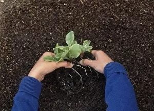
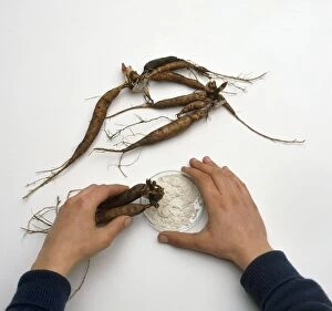
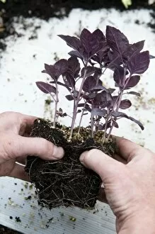
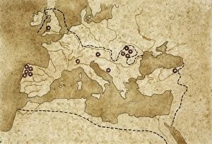
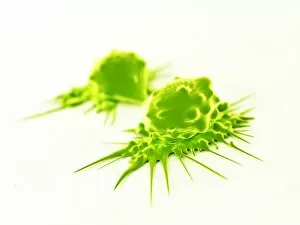
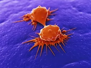
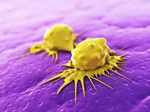
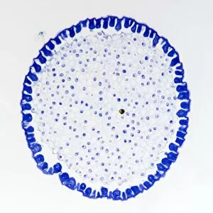
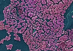
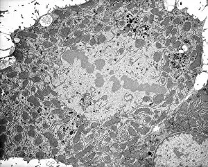
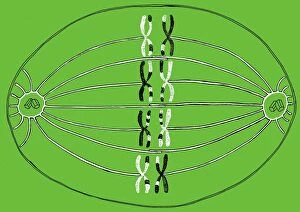
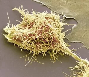
"Exploring the Intricate Process of Dividing: From Horses to Microorganisms" In a tranquil stable, two horses stand side by side while a small terrier curiously observes their peaceful coexistence. Under the lens of a light micrograph, we witness the mesmerizing beauty of mitosis as cells divide and multiply, creating new life. Delving deeper into the microscopic world, dividing yeast cells come alive in stunning detail through scanning electron microscopy (SEM). The resilient E. Coli bacteria demonstrate their remarkable ability to divide and propagate, showcasing nature's intricate mechanisms on a minuscule scale. Fluorescent micrographs unveil the captivating process of cell division, where vibrant hues highlight each step towards growth and renewal. Sketches depicting St Kilda in Western Hebrides transport us back in time when human settlements were divided by vast landscapes yet connected through shared experiences. Witnessing embryo development 24-36 hours after fertilization reveals how life begins with rapid cell divisions that lay the foundation for future growth and complexity. Whether it is within organisms or across species boundaries, dividing cells remind us of nature's constant drive for renewal and adaptation. Another glimpse into fluorescent micrographs captures the awe-inspiring dance of cell division as it unfolds within different tissues and organisms alike. Stem cells take center stage under scanning electron microscopy (SEM), displaying their potential to differentiate into various specialized cell types—a testament to our body's regenerative capabilities. Even at such minute scales, E. coli bacterium displays its resilience through transmission electron microscopy (TEM), revealing its ability to divide despite challenging conditions—nature's ultimate survivor.