Dividing Collection (page 8)

"Exploring the Intricate Process of Dividing: From Horses to Microorganisms" In a tranquil stable

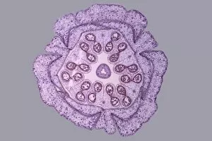
Background imageDividing Collection: Lily flower bud, light micrograph

Lily flower bud, light micrograph
Lily flower bud. Light micrograph of a section through a young flower bud from a lily (Lilium sp.), showing pollen cells (circles) dividing within the anthers (inner ring)
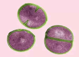
Background imageDividing Collection: Enterococcus faecalis dividing, TEM

Enterococcus faecalis dividing, TEM
HPA Colindale 12/01/12 12:30:37 15000 7.0 80.0 Imaging Enterococcus faecalis 1.5 AmMob plus wash -170.96 657.83 . XpixCal=1.242 YpixCal=1.242 Unit=nm ##fv3

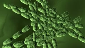
Background imageDividing Collection: Dividing bacteria, computer artwork

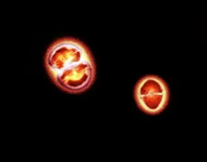
Dividing bacteria, computer artwork
Bacteria dividing, computer artwork

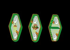
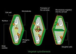
Background imageDividing Collection: Cytokinesis, artwork

Cytokinesis, artwork
Cytokinesis. Artwork showing the stage of cell division that involves the splitting of the cell cytoplasm between two daughter cells

Background imageDividing Collection: Cell division, artwork

Cell division, artwork
Cell division. Computer artwork of an animal cell undergoing mitosis (nuclear division) and cytokinesis (cell division) to form two identical daughter cells (bottom)

Background imageDividing Collection: Cytokinesis, diagram

Cytokinesis, diagram
Cytokinesis. Diagram showing the stage of cell division that involves the splitting of the cell cytoplasm between two daughter cells

Background imageDividing Collection: Bacterial endospore formation, diagram

Bacterial endospore formation, diagram. The initial stage is at upper left, following the arrow to a ninth stage at lower right

Background imageDividing Collection: Cell division, conceptual artwork

Cell division, conceptual artwork
cell division

Background imageDividing Collection: Bacterial replication, artwork

Bacterial replication, artwork
Bacterial replication. Computer artwork showing a single bacterium dividing to form two identical daughter cells. This process is a form of asexual reproduction and is known as binary fission

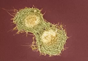
Background imageDividing Collection: Cell division, SEM

Cell division, SEM
Cell division. Coloured scanning electron micrograph (SEM) of two animal cells during cytokinesis (cell division). Cytokinesis occurs after nuclear division (mitosis)

Background imageDividing Collection: Young stem, light micrograph

Young stem, light micrograph. Longitudinal section through a young stem showing an intercalary meristem. Intercalary meristems are found at the base of grass leaves

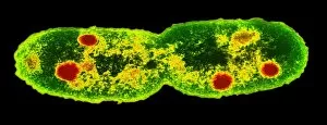
Background imageDividing Collection: TEM of dividing E. coli bacterium

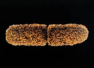
TEM of dividing E. coli bacterium
False-colour transmission electron micrograph of the bacterium Escherichia coli, a normal inhabitant of the human intestine

Background imageDividing Collection: False-colour TEM of root cell division

False-colour TEM of root cell division
False-colour transmission electron micrograph of a late stage of cell division in the root tip cell (meristem) of Zea mays, maize

Background imageDividing Collection: Stenotrophomonas sp. bacteria, TEM

Stenotrophomonas sp. bacteria, TEM
Stenotrophomonas maltophilia bacteria. Coloured transmission electron micrograph (TEM) of a sect- ion through Stenotrophomonas maltophilia bacteria. They are seen on a fractal background

Background imageDividing Collection: Mycobacterium dividing, TEM

Mycobacterium dividing, TEM
Mycobacterium dividing. Coloured transmission electron micrograph (TEM) of a Mycobacterium sp. bacterium undergoing binary fission

Background imageDividing Collection: Chlamydia bacteria, TEM

Chlamydia bacteria, TEM
Chlamydia bacteria. Coloured transmission electron micrograph (TEM) of Chlamydia sp. bacteria (small blue spheres) in a human cell (large blue sphere)

Background imageDividing Collection: Cryptococcus cell dividing, TEM

Cryptococcus cell dividing, TEM
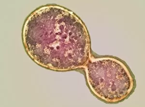
Cryptococcus cell dividing. Coloured transmission electron micrograph (TEM) of the asexual budding (lower right) of a Cyptococcus sp. yeast cell

Background imageDividing Collection: TEM of Staphylococcus aureus bacteria

TEM of Staphylococcus aureus bacteria
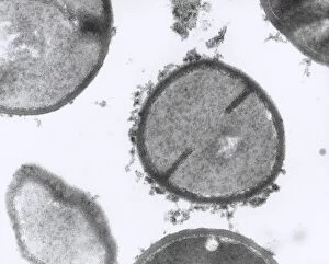
Staphylococcus bacteria. Transmission electron micrograph (TEM) of sectioned Staphylococcus aureus bacteria. Staphylococcus aureus are Gram- positive coccal (spherical) bacteria

Background imageDividing Collection: Gut bacterium reproducing, TEM

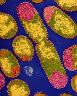
Gut bacterium reproducing, TEM
Gut bacterium reproducing. Coloured transmission electron micrograph (TEM) of a single bacterium dividing into two identical clone daughter bacteria

Background imageDividing Collection: Coloured TEM of E. coli bacteria dividing

Coloured TEM of E. coli bacteria dividing
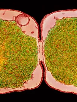
E. Coli bacteria dividing. Transmission electron micrograph of the division zone between two new Escherichia coli bacteria showing the cells undergoing binary fission

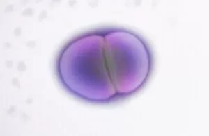
Background imageDividing Collection: Moraxella catarrhalis bacterium dividing

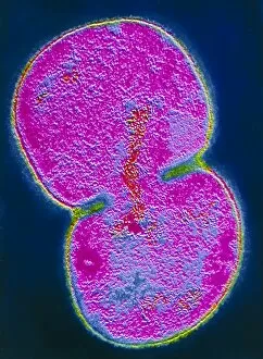
Moraxella catarrhalis bacterium dividing

Background imageDividing Collection: Cell division in Salmonella bacterium

Cell division in Salmonella bacterium
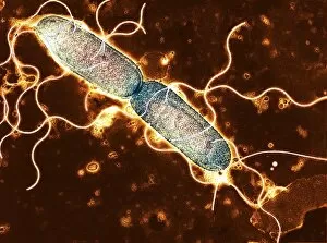
Salmonella cell dividing. Coloured Transmission electron micrograph of the bacterium Salmonella typhimurium undergoing division by binary fission

Background imageDividing Collection: Dividing cell in maize root tip

Dividing cell in maize root tip

Background imageDividing Collection: Mathematics

Mathematics. Computer artwork of numbers and mathematical symbols

Background imageDividing Collection: Artwork of dividing E. coli bacterium

Artwork of dividing E. coli bacterium
Artwork of the bacterium Escherichia coli during cell division. It belongs to the genus of Gram- negative rod-shaped bacteria and is generally motile

Background imageDividing Collection: Staphylococcus aureus bacteria dividing

Staphylococcus aureus bacteria dividing
Staphylococcus bacteria. Coloured transmission electron micrograph (TEM) of Staphylococcus aureus bacteria (light blue), some undergoing cell division

Background imageDividing Collection: MRSA bacteria

MRSA bacteria. Coloured transmission electron micrograph (TEM) of Methicillin-resistant Staphylococcus aureus (MRSA), a Gram-positive, round (coccus) bacterium

Background imageDividing Collection: MRSA bacteria, TEM

MRSA bacteria, TEM
MRSA bacteria. Coloured transmission electron micrograph (TEM) of methicillin-resistant Staphylococcus aureus (MRSA), a gram-positive, round (coccus) bacterium

Background imageDividing Collection: Streptococcus oral bacteria, TEM

Streptococcus oral bacteria, TEM
Streptococcus oral bacteria. Coloured transmission electron micrograph (TEM) of two Streptococcus sanguinis bacteria. They are undergoing nuclear division (mitosis)

Background imageDividing Collection: Kirkpatrick Fleming Poorhouse, Dumfriesshire, Scotland

Kirkpatrick Fleming Poorhouse, Dumfriesshire, Scotland
View of the Kirkpatrick Fleming Combination poorhouse, Dumfriesshire, Scotland. The image is caption " Rest for the Weary" -- the poorhouse mostly catered for the elderly and the bedridden

Background imageDividing Collection: Union Infirmary, Wigan, Lancashire

Union Infirmary, Wigan, Lancashire
The Wigan Union infirmary, Upholland Road, Wigan - an unusual composite of two photographs. The infirmary, later known as Billinge Hospital

Background imageDividing Collection: Egypt - Tourists hassled by locals

Egypt - Tourists hassled by locals
Humourous postcard, depicting the hardships endured by European Tourists in Egypt, cornered by a vast horde of helpful Egyptians who are aggressively " Dividing the Spoils" !

Background imageDividing Collection: Ligue Catholique

Ligue Catholique
Allegorical depiction of the Catholic League - showing the serpent-headed League dividing its loyalty to the king and elsewhere

Background imageDividing Collection: Fault in the landscape caused by continental drift between North American

Fault in the landscape caused by continental drift between North American and Eurasian tectonic plates at Thingvellir National Park near Reykjavik, Iceland, Polar Regions

Background imageDividing Collection: Colorado River dividing California and Arizona

Colorado River dividing California and Arizona, near Parker, Arizona, United States of America, North America

Background imageDividing Collection: Snow covered bridges across Vltava River dividing Old Town from Mala Strana suburbs in winter

Snow covered bridges across Vltava River dividing Old Town from Mala Strana suburbs in winter twilight, Mala Strana, Prague, Czech Republic, Europe

Background imageDividing Collection: The Great Wall of China, Mutianyu, China

The Great Wall of China, Mutianyu, China

Background imageDividing Collection: Staphylococcus aureus dividing, TEM

Staphylococcus aureus dividing, TEM
Staphylococcus aureus bacteria dividing, coloured transmission electron micrograph. Atlower left is a single bacterium, the bacterium atupper centre is starting to divide

Background imageDividing Collection: Oral bacteria, TEM

Oral bacteria, TEM
Oral bacteria. Computer-enhanced colouredtransmission electron micrograph (TEM) of Porphyromonas gingivalis bacteria (formerlynamed Bacteroides gingivalis )

Background imageDividing Collection: Listeria monocytogenes bacteria, TEM

Listeria monocytogenes bacteria, TEM
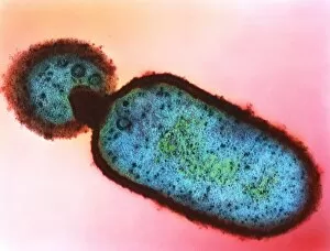
Listeria bacteria. Coloured transmissionelectron micrograph (TEM) of a Listeria monocytogenes bacterium (centre to bottomright). It is dividing by a process of unequalcell growth known as budding

Background imageDividing Collection: Napoleon / Pitt / World / 1805

Napoleon / Pitt / World / 1805
The Plumb-pudding in danger Napoleon and Pitt carve up the world at dinner


For sale as Licensed Images

Choose your image, Select your licence and Download the media

"Exploring the Intricate Process of Dividing: From Horses to Microorganisms" In a tranquil stable, two horses stand side by side while a small terrier curiously observes their peaceful coexistence. Under the lens of a light micrograph, we witness the mesmerizing beauty of mitosis as cells divide and multiply, creating new life. Delving deeper into the microscopic world, dividing yeast cells come alive in stunning detail through scanning electron microscopy (SEM). The resilient E. Coli bacteria demonstrate their remarkable ability to divide and propagate, showcasing nature's intricate mechanisms on a minuscule scale. Fluorescent micrographs unveil the captivating process of cell division, where vibrant hues highlight each step towards growth and renewal. Sketches depicting St Kilda in Western Hebrides transport us back in time when human settlements were divided by vast landscapes yet connected through shared experiences. Witnessing embryo development 24-36 hours after fertilization reveals how life begins with rapid cell divisions that lay the foundation for future growth and complexity. Whether it is within organisms or across species boundaries, dividing cells remind us of nature's constant drive for renewal and adaptation. Another glimpse into fluorescent micrographs captures the awe-inspiring dance of cell division as it unfolds within different tissues and organisms alike. Stem cells take center stage under scanning electron microscopy (SEM), displaying their potential to differentiate into various specialized cell types—a testament to our body's regenerative capabilities. Even at such minute scales, E. coli bacterium displays its resilience through transmission electron microscopy (TEM), revealing its ability to divide despite challenging conditions—nature's ultimate survivor.