
Electron Collection (#2)
"The Electron: Unveiling the Mysteries of Particle Physics and Beyond" In the vast realm of particle physics


For sale as Licensed Images
Choose your image, Select your licence and Download the media
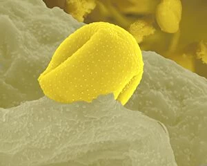
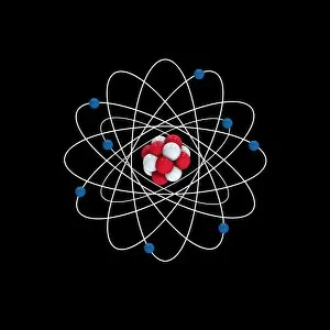
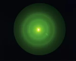
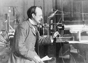
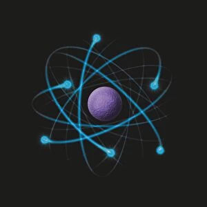
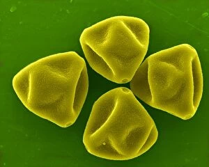
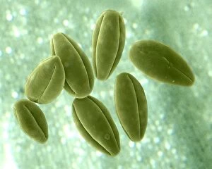
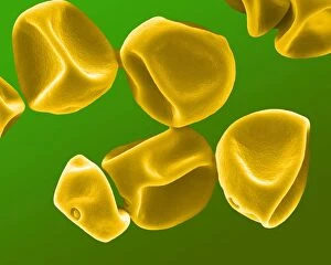
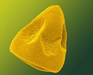
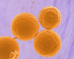
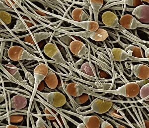
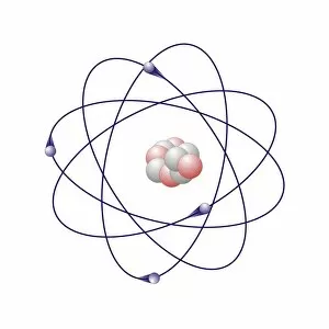
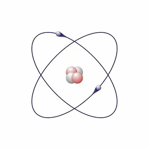
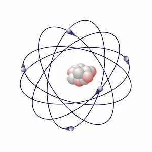
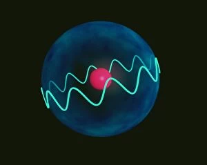
"The Electron: Unveiling the Mysteries of Particle Physics and Beyond" In the vast realm of particle physics, the electron stands as a fundamental building block that has captivated scientists for decades. As we delve into its enigmatic nature, we are greeted with awe-inspiring visuals that shed light on its intricate properties. One such image is the mesmerizing bubble chamber photo capturing the decay of a sigma particle. This snapshot reveals the hidden dance between particles, unraveling their secrets within complex equations adorning scientific papers. Artwork depicting particle physics experiments further immerses us in this captivating world. It serves as a visual testament to human curiosity and our relentless pursuit of knowledge. Among these illustrations, Niels Bohr's caricature reminds us of his groundbreaking contributions to atomic theory. Nuclear fission artwork showcases humanity's quest for harnessing immense energy from splitting atoms—an achievement that forever altered our understanding of power generation and weaponry. The Higgs boson, often referred to as "the God particle, " takes center stage in another remarkable artwork. Its discovery revolutionized our comprehension of mass and solidified our understanding of how particles acquire their weight. Beyond subatomic realms lie unexpected connections—like Simulium damnosum, also known as Simulian blackfly. These tiny creatures possess an intriguing link to electrons through their unique ability to transmit diseases like river blindness—a reminder that science encompasses all facets of life. Delving deeper into atomic structures brings forth stunning artwork showcasing intricate arrangements resembling delicate lacework or snail teeth—a testament to nature's elegance even at microscopic scales. As we revisit those familiar equations describing electron structure within helium atoms, we marvel at how these minuscule entities shape everything around us—the foundation upon which matter is built. The electron remains an ever-present force shaping our world—from powering electronic devices to enabling chemical reactions essential for life itself. Its significance cannot be overstated; it embodies both simplicity and complexity, a paradox that continues to intrigue and inspire scientists worldwide.