Electron Collection (#7)

"The Electron: Unveiling the Mysteries of Particle Physics and Beyond" In the vast realm of particle physics

Background imageElectron Collection: 19th Century Crookes Tube

19th Century Crookes Tube. Invented by William Crookes (1832-1919) in the late 19th century this apparatus was used to investigate the path taken by electrons, or cathode rays as they were known

Background imageElectron Collection: 19th Century Crookes Tubes

19th Century Crookes Tubes. Invented by William Crookes (1832-1919) in the late 19th century this apparatus was used to investigate the path taken by electrons, or cathode rays as they were known

Background imageElectron Collection: Hydrogen atom, conceptual model C013 / 5605

Hydrogen atom, conceptual model C013 / 5605
Hydrogen atom, conceptual model. Computer artwork representing the atomic structure of hydrogen. Hydrogen has one proton and one neutron (large spheres) in its nucleus (large circle, centre)

Background imageElectron Collection: Helium atom, conceptual model C013 / 5600

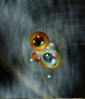
Helium atom, conceptual model C013 / 5600
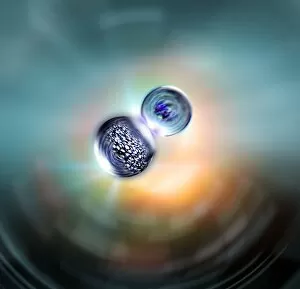
Helium atom, conceptual model. Computer artwork representing the atomic structure of helium. Helium has two protons and two neutrons (large spheres) in its nucleus (faint circle, centre)

Background imageElectron Collection: Helium atom, conceptual model C013 / 5601

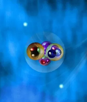
Helium atom, conceptual model C013 / 5601
Helium atom, conceptual model. Computer artwork representing the atomic structure of helium. Helium has two protons and two neutrons (large spheres) in its nucleus (faint circle, centre)

Background imageElectron Collection: Atomic interactions, conceptual image C013 / 5595

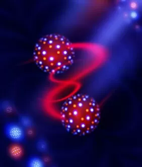
Atomic interactions, conceptual image C013 / 5595
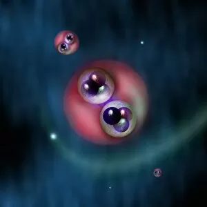
Atomic interactions, conceptual image. Computer artwork representing the interactions between atomic and sub-atomic particles

Background imageElectron Collection: Photon emission, artwork

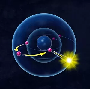
Photon emission, artwork
Photon emission. Computer artwork of an atom (large sphere) emitting a photon (yellow). The atom consists of a nucleus (blue, centre), which contains neutrons and protons (not shown)

Background imageElectron Collection: Calcium carbonate, SEM C017 / 8495

Calcium carbonate, SEM C017 / 8495
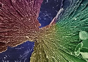
False-coloured Scanning Electron Micrograph (SEM) of microscopic crystals of calcium carbonate. Calcium carbonate has many polymorphs (different shaped) crystal forms

Background imageElectron Collection: Calcium carbonate, SEM C017 / 8497

Calcium carbonate, SEM C017 / 8497
False-coloured Scanning Electron Micrograph (SEM) of microscopic crystals of calcium carbonate. Calcium carbonate has many polymorphs (different shaped) crystal forms

Background imageElectron Collection: Calcium carbonate, SEM C017 / 8491

Calcium carbonate, SEM C017 / 8491
False-coloured Scanning Electron Micrograph (SEM) of microscopic crystals of calcium carbonate. Calcium carbonate has many polymorphs (different shaped) crystal forms

Background imageElectron Collection: Particles, conceptual artwork C013 / 5639

Particles, conceptual artwork C013 / 5639
Particles, conceptual computer artwork

Background imageElectron Collection: Quantum states, conceptual artwork C013 / 5630

Quantum states, conceptual artwork C013 / 5630
Quantum states, conceptual artwork. In physics, a quantum state is a set of mathematical variables that fully describes a quantum system

Background imageElectron Collection: Particles, conceptual artwork C013 / 5626

Particles, conceptual artwork C013 / 5626
Particles, conceptual computer artwork

Background imageElectron Collection: Particles, conceptual artwork C013 / 5627

Particles, conceptual artwork C013 / 5627
Particles, conceptual computer artwork

Background imageElectron Collection: Structure of matter, artwork C017 / 8029

Structure of matter, artwork C017 / 8029
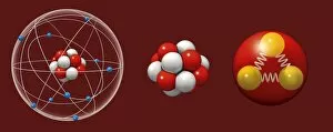
Structure of matter. Computer artwork representing the Standard Model of particle physics. Shown here are an atom (left) composed of electrons (blue) orbiting a central nucleus

Background imageElectron Collection: Hydrogen atoms, conceptual model C013 / 5606

Hydrogen atoms, conceptual model C013 / 5606
Hydrogen atoms, conceptual model. Computer artwork representing the structure of hydrogen atoms. Each atom has one proton and one neutron (large spheres) in its nucleus (pink)

Background imageElectron Collection: Researcher with particle accelerator C015 / 6204

Researcher with particle accelerator C015 / 6204
Materials science. The U-28 electron accelerator being prepared for use in a materials science experiment. U-28 is based at the National Research Nuclear University in Moscow, Russia

Background imageElectron Collection: Crookes cathode ray tube, 1880s C016 / 3666

Crookes cathode ray tube, 1880s C016 / 3666
Crookes tube. A Crookes tube, used to create cathode rays, made in the 1880s. This is a tube containing a near vacuum. A high voltage is applied across the two electrodes

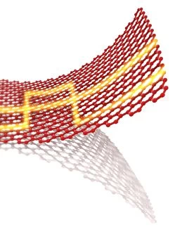
Background imageElectron Collection: Graphene conductivity, conceptual image C013 / 8897

Graphene conductivity, conceptual image C013 / 8897
Graphene conductivity, conceptual image. Computer artwork of a graphene sheet with electricity (yellow) passing through it. Graphene is a single layer of graphite

Background imageElectron Collection: Archaea (Methanosarcina sp. ) SEM C013 / 7189

Archaea (Methanosarcina sp. ) SEM C013 / 7189
Archaea (Methanosarcina sp.) scanning electron micrograph (SEM). These primitive archaebacteria have unusual cell walls and membranes and are methane producers

Background imageElectron Collection: Archaea (Methanosarcina sp. ) SEM C013 / 7190

Archaea (Methanosarcina sp. ) SEM C013 / 7190
Archaea (Methanosarcina sp.) scanning electron micrograph (SEM). These primitive archaebacteria have unusual cell walls and membranes and are methane producers

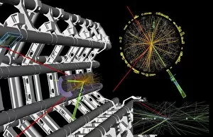
Background imageElectron Collection: Higgs boson research, ATLAS detector C013 / 6893

Higgs boson research, ATLAS detector C013 / 6893
Higgs boson research. 3D computer graphic showing one of the numerous particle collision events recorded during the search for the Higgs boson

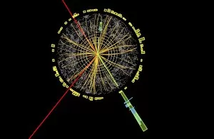
Background imageElectron Collection: Higgs boson research, ATLAS detector C013 / 6894

Higgs boson research, ATLAS detector C013 / 6894
Higgs boson research. Graphic of a transverse section through a detector showing one of the numerous particle collision events recorded during the search for the Higgs boson

Background imageElectron Collection: Higgs boson research, ATLAS detector C013 / 6889

Higgs boson research, ATLAS detector C013 / 6889
Higgs boson research. Graphic of a transverse section through a detector showing one of the numerous particle collision events recorded during the search for the Higgs boson

Background imageElectron Collection: Higgs boson research, ATLAS detector C013 / 6888

Higgs boson research, ATLAS detector C013 / 6888
Higgs boson research. 3D computer graphic showing one of the numerous particle collision events recorded during the search for the Higgs boson

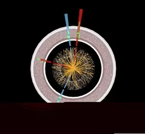
Background imageElectron Collection: Higgs boson research, CMS detector C013 / 6884

Higgs boson research, CMS detector C013 / 6884
Higgs boson research. Graphic of a longitudinal section through a detector showing a collision event recorded during the search for the Higgs boson

Background imageElectron Collection: Higgs boson research, CMS detector C013 / 6882

Higgs boson research, CMS detector C013 / 6882
Higgs boson research. 3D computer graphic showing one of the numerous particle collision events recorded during the search for the Higgs boson

Background imageElectron Collection: Blitz in London -- training office workers, WW2

Blitz in London -- training office workers, WW2
Blitz in London -- LFS personnel training office workers in firefighting, 7 June 1941, with a diagram of a typical kilo magnesium electron incendiary bomb pinned to a blackboard

Background imageElectron Collection: Pieris rapae, small white

Pieris rapae, small white
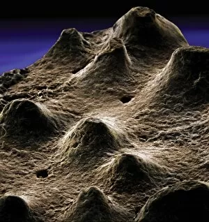
SEM image of the wing of a small white butterfly

Background imageElectron Collection: Aglais urticae, small tortoiseshell butterfly

Aglais urticae, small tortoiseshell butterfly
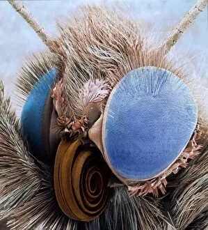
Scanning electron microscope image showing the head of a small tortoiseshell butterfly (x 25 on a standard 9cm wide print). This image has been coloured artificially by computer

Background imageElectron Collection: Dinosaur eggshell

Dinosaur eggshell
Scanning electron microscope image on display in the Darwin Centre

Background imageElectron Collection: Ventral surface of a mite from the prostigmatic species

Ventral surface of a mite from the prostigmatic species
Scanning electron microscope image displayed on the glass screens in the Darwin Centre, at the Natural History Museum, London

Background imageElectron Collection: Diplopoda sp. plate millipede

Diplopoda sp. plate millipede
Scanning electron microscope image of a lateral view of the head of a plate millipede. Image displayed on the glass screens in the Darwin Centre, at the Natural History Museum, London

Background imageElectron Collection: Trypanosomes

Trypanosomes
Scanning electron microscope image showing a trypanosoma blood smear. They have proved to be of great interest as they have evolved very differently to other better studied organisms

Background imageElectron Collection: Atta cethalotes, leaf-cutter ant

Atta cethalotes, leaf-cutter ant
Scanning electron microscope image of a leaf-cutter ant displayed in the Darwin Centre, at the Natural History Museum, London

Background imageElectron Collection: Small fly, species unknown

Small fly, species unknown
Scanning electron microscope (SEM) of a fly head. Image displayed on the glass screens in the Darwin Centre, at the Natural History Museum, London

Background imageElectron Collection: Woodlouse antenna

Woodlouse antenna
Scanning Electron Microscope (SEM) image of woodlouse antenna

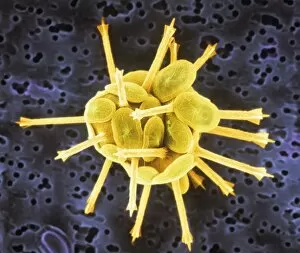
Background imageElectron Collection: Amphitetras, diatom

Amphitetras, diatom
Scanning electron microscope (SEM) image showing the diatom Amphitetras with its ornate silica shell (x5000 on a standard 9 cm wide print). Coloured artificially by computer

Background imageElectron Collection: Leptoglossis ferreyraei

Leptoglossis ferreyraei
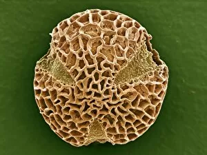
A pollen grain of Leptoglossis ferreyraei (polar view) from the family Solanaceae, the tomato family

Background imageElectron Collection: Leptoglossis lomana

Leptoglossis lomana
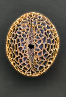
A pollen grain of the Leptoglossis lomana (polar view) from the family Solanacea, the tomato family

Background imageElectron Collection: Salticus senecus, zebra jumping spider

Salticus senecus, zebra jumping spider
Scanning electron microscope image of a zebra jumping spider from the UK (x 35). Note the two large eyes that give them excellent binoular vision. Coloured artificially on computer

Background imageElectron Collection: Gold in unspecified mineral

Gold in unspecified mineral
Scanning electron microscope image of an elemental map showing the distribution of gold (Au) in mineral samples

Background imageElectron Collection: Goyazite

Goyazite
Scanning electron microscope image of the energy-dispersive X-ray spectrum of the mineral goyazite, obtained using Link AN10000 analysis system

Background imageElectron Collection: Coccoliths

Coccoliths
Scanning electron microscope (SEM) image of coccoliths, these are the limestone scales surrounding the marine phytoplankton coccolithophores

Background imageElectron Collection: Sitophilus granarius, grain weevil

Sitophilus granarius, grain weevil
Scanning electron microscope image of a grain weevil (x 50). Note the elongated snout or rostrum, with the chewing mouthparts at the end. These weevils cannot fly. Artificially coloured by computer

Background imageElectron Collection: Velcro

Velcro
A trademarked name for a fastening tape made up of a strip of nylon with a surface of minute hooks, that fasten to another strip with a surface of uncut pile. A SEM image

Background imageElectron Collection: Amblyomma sp. hard backed tick

Amblyomma sp. hard backed tick
Scanning electron microscope view of a hard backed tick from the family Ixodidae. Coloured artificially on computer

Background imageElectron Collection: Copper in unspecified mineral

Copper in unspecified mineral
Scanning electron microscope image of an elemental map showing the distribution of copper (Cu) in mineral samples


For sale as Licensed Images

Choose your image, Select your licence and Download the media

"The Electron: Unveiling the Mysteries of Particle Physics and Beyond" In the vast realm of particle physics, the electron stands as a fundamental building block that has captivated scientists for decades. As we delve into its enigmatic nature, we are greeted with awe-inspiring visuals that shed light on its intricate properties. One such image is the mesmerizing bubble chamber photo capturing the decay of a sigma particle. This snapshot reveals the hidden dance between particles, unraveling their secrets within complex equations adorning scientific papers. Artwork depicting particle physics experiments further immerses us in this captivating world. It serves as a visual testament to human curiosity and our relentless pursuit of knowledge. Among these illustrations, Niels Bohr's caricature reminds us of his groundbreaking contributions to atomic theory. Nuclear fission artwork showcases humanity's quest for harnessing immense energy from splitting atoms—an achievement that forever altered our understanding of power generation and weaponry. The Higgs boson, often referred to as "the God particle, " takes center stage in another remarkable artwork. Its discovery revolutionized our comprehension of mass and solidified our understanding of how particles acquire their weight. Beyond subatomic realms lie unexpected connections—like Simulium damnosum, also known as Simulian blackfly. These tiny creatures possess an intriguing link to electrons through their unique ability to transmit diseases like river blindness—a reminder that science encompasses all facets of life. Delving deeper into atomic structures brings forth stunning artwork showcasing intricate arrangements resembling delicate lacework or snail teeth—a testament to nature's elegance even at microscopic scales. As we revisit those familiar equations describing electron structure within helium atoms, we marvel at how these minuscule entities shape everything around us—the foundation upon which matter is built. The electron remains an ever-present force shaping our world—from powering electronic devices to enabling chemical reactions essential for life itself. Its significance cannot be overstated; it embodies both simplicity and complexity, a paradox that continues to intrigue and inspire scientists worldwide.