False Coloured Collection (page 8)

"Revealing the Unseen: Exploring the World Through False Colors" Step back in time to medieval alchemy

Background imageFalse Coloured Collection: Moth orchid (Phalaenopsis orchidaceae), X-ray

Moth orchid (Phalaenopsis orchidaceae), X-ray
Moth orchid (Phalaenopsis orchidaceae), coloured X-ray

Background imageFalse Coloured Collection: Wheat, X-ray

Wheat, X-ray
Wheat, coloured X-ray

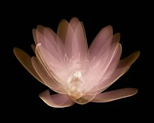
Background imageFalse Coloured Collection: Water lily (Nymphaea alba), X-ray

Water lily (Nymphaea alba), X-ray
Water lily (Nymphaea alba), coloured X-ray

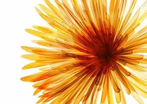
Background imageFalse Coloured Collection: Chrysanthemum, X-ray

Chrysanthemum, X-ray
Chrysanthemum, coloured X-ray

Background imageFalse Coloured Collection: Pomegranates, X-ray

Pomegranates, X-ray
Pomegranates, coloured X-ray

Background imageFalse Coloured Collection: Blue Danube (Azalea japonica), X-ray

Blue Danube (Azalea japonica), X-ray
Blue Danube (Azalea japonica), coloured X-ray

Background imageFalse Coloured Collection: Broccoli (Brassica oleracea), X-ray

Broccoli (Brassica oleracea), X-ray
Broccoli (Brassica oleracea), coloured X-ray

Background imageFalse Coloured Collection: Thistle, X-ray

Thistle, X-ray
Thistle, coloured X-ray

Background imageFalse Coloured Collection: Begonia (Begonia crenata) leaf, X-ray

Begonia (Begonia crenata) leaf, X-ray
Begonia (Begonia crenata) leaf, coloured X-ray

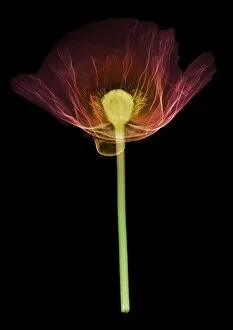
Background imageFalse Coloured Collection: Poppy (Papaver orientalis), X-ray

Poppy (Papaver orientalis), X-ray
Poppy (Papaver orientalis), coloured X-ray

Background imageFalse Coloured Collection: Lavender, X-ray

Lavender, X-ray
Lavender, coloured X-ray

Background imageFalse Coloured Collection: Pink rose (Rosa centifolia), X-ray

Pink rose (Rosa centifolia), X-ray
Pink rose (Rosa centifolia), coloured X-ray

Background imageFalse Coloured Collection: Peony (Paeonia suffruticosa), X-ray

Peony (Paeonia suffruticosa), X-ray
Peony (Paeonia suffruticosa), coloured X-ray

Background imageFalse Coloured Collection: Water lily, X-ray

Water lily, X-ray
Water lily, coloured X-ray

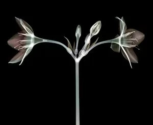
Background imageFalse Coloured Collection: Eucharis lily, X-ray

Eucharis lily, X-ray
Eucharis lily, coloured X-ray

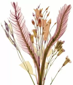
Background imageFalse Coloured Collection: Long leaves and pastel flower buds, X-ray

Long leaves and pastel flower buds, X-ray
Long leaves and pastel flower buds, coloured X-ray

Background imageFalse Coloured Collection: Artichoke leaf, X-ray

Artichoke leaf, X-ray
Artichoke leaf, coloured X-ray

Background imageFalse Coloured Collection: Patchouli leaves (Pogostemon cablin), X-ray

Patchouli leaves (Pogostemon cablin), X-ray
Patchouli leaves (Pogostemon cablin), coloured X-ray

Background imageFalse Coloured Collection: Pink water lily (Nymphaea sp. ), X-ray

Pink water lily (Nymphaea sp. ), X-ray
Pink water lily (Nymphaea sp.), coloured X-ray

Background imageFalse Coloured Collection: Guelder rose leaf (Viburnum opulus), X-ray

Guelder rose leaf (Viburnum opulus), X-ray
Guelder rose leaf (Viburnum opulus), coloured X-ray

Background imageFalse Coloured Collection: Pink and violet lily blossom, X-ray

Pink and violet lily blossom, X-ray
Pink and violet lily blossom, coloured X-ray

Background imageFalse Coloured Collection: Artificial green fibre optic festive tree, X-ray

Artificial green fibre optic festive tree, X-ray
Artificial green fibre optic festive tree, coloured X-ray

Background imageFalse Coloured Collection: Primrose (Primula vulgaris), X-ray

Primrose (Primula vulgaris), X-ray

Background imageFalse Coloured Collection: Bundle of flowers (Dianthus sp. ), X-ray

Bundle of flowers (Dianthus sp. ), X-ray
Bundle of flowers (Dianthus sp.), coloured X-ray

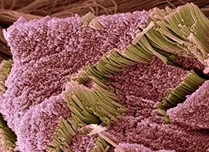
Background imageFalse Coloured Collection: Tendon, SEM

Tendon, SEM
Tendon, coloured scanning electron micrograph (SEM), showing bundles of collagen fibres. The parallel alignment of the fibres make tendons inelastic but flexible. Tendons attach muscle to bone

Background imageFalse Coloured Collection: Brochosomes, SEM

Brochosomes, SEM
Brochosomes, coloured scanning electron micrograph (SEM). Brochosomes are intricately structured microscopic granules secreted by leafhoppers

Background imageFalse Coloured Collection: Verbena pollen, SEM

Verbena pollen, SEM
Verbena pollen. Coloured scanning electron micrograph (SEM) of pollen grains from verbena bonariensis. Verbena bonariensis is a tall, slender-stemmed perennial

Background imageFalse Coloured Collection: Diatoms, SEM

Diatoms, SEM
Diatoms. Coloured scanning electron micrograph (SEM) of fresh water centric diatom frustules (skeleton), Diatoms are a type of algae (Chromophyta, Bacillariophyceae)

Background imageFalse Coloured Collection: Convolvulus pollen grains, SEM

Convolvulus pollen grains, SEM
Convolvulus pollen grains. Coloured scanning electron micrograph (SEM) of pollen grains from a convolvulus flower. Convolvulus is a genus of about 200 to 250 species of flowering plants in

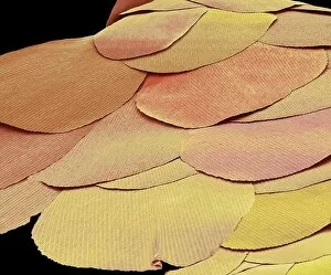
Background imageFalse Coloured Collection: Silverfish scales, SEM

Silverfish scales, SEM
Silverfish scales (Lepisma saccharina). Coloured scanning electron micrograph (SEM) of scales from a silverfish. The silverfish is a primitive insect that has remained unchanged for millions of

Background imageFalse Coloured Collection: Water lily pollen grains, SEM

Water lily pollen grains, SEM
Water lily pollen grains. Coloured scanning electron micrograph (SEM) of pollen grains from a water lily flower. Nymphaeaceae is a family of flowering plants, commonly called water lilies

Background imageFalse Coloured Collection: Human hair, sem

Human hair, sem
Human hair (Caucasian, brunette), coloured scanning electron micrograph (SEM). The outer layer of hair (the cuticle) has overlapping scales of keratin

Background imageFalse Coloured Collection: Shed claw from a cat, SEM

Shed claw from a cat, SEM
Cats claw, coloured scanning electron micrograph (SEM). This is the shed outer layer of the claw. Domestic cats typically shed the outer layers of their claws, which grow from the inside

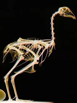
Background imageFalse Coloured Collection: Ostrich skeleton

Ostrich skeleton. Coloured x-ray of an ostrich (Struthio camelus). This bird is native to Africa and it is the largest species of bird

Background imageFalse Coloured Collection: Phagocytosis of fungus spores, SEM

Phagocytosis of fungus spores, SEM
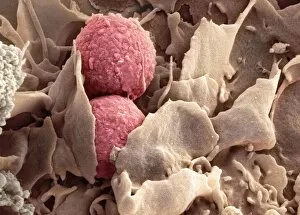
Phagocytosis of fungus spores. Coloured scanning electron micrograph (SEM) of spores from the fungus Aspergillus fumigatus (red) being phagocytosed (engulfed and destroyed) by a dendritic cell (brown)

Background imageFalse Coloured Collection: Broad bean pollen, SEM

Broad bean pollen, SEM
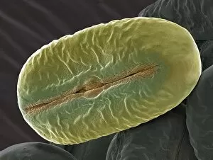
Broad bean pollen. Coloured scanning electron micrograph (SEM) of pollen grains from a Vicia faba plant. Magnification: x2000 when printed 10 centimetres wide

Background imageFalse Coloured Collection: Plant cell mitosis, light micrograph

Plant cell mitosis, light micrograph
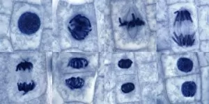
Plant cell mitosis. Light micrograph of root tip cells from an onion (Allium sp.) undergoing cell division (mitosis). From top left to bottom right

Background imageFalse Coloured Collection: Deformed sperm cell, SEM

Deformed sperm cell, SEM
Deformed sperm cell. Coloured scanning electron micrograph (SEM) of double-tailed sperm cell, or spermatozoon. A sperm cells motility may be seriously affected by having multiple heavy tails

Background imageFalse Coloured Collection: Snake head, SEM

Snake head, SEM
Snake (suborder Serpentes) head, coloured scanning electron micrograph (SEM). The snakes forked tongue (right) is used to detect prey by picking up scent particles from the air

Background imageFalse Coloured Collection: Chloroplast, SEM

Chloroplast, SEM
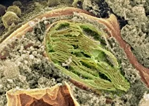
Chloroplast. Coloured scanning electron micrograph (SEM) of a section through a plant cell, showing a fractured chloroplast (green)

Background imageFalse Coloured Collection: Inner ear hairs, SEM

Inner ear hairs, SEM
Inner ear hairs. Coloured scanning electron micrograph (SEM) of stereocilia, sensory hair-like projections, from hair cells of the cochlea of the inner ear

Background imageFalse Coloured Collection: Yeast cells, SEM

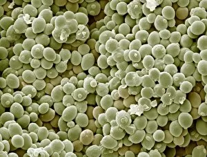
Yeast cells, SEM
Yeast cells. Coloured scanning electron micrograph (SEM) of cells of bakers yeast (Saccharomyces cerevisiae) from part of a dried, commercial yeast pellet

Background imageFalse Coloured Collection: Electrical sewing machine, 1900

Electrical sewing machine, 1900
Electrical sewing machine. Historical artwork of a woman using a 19th-century electrical sewing machine. She is using her feet to operate a pedal that moves an electrical contact over a rheostat

Background imageFalse Coloured Collection: Monkey skeleton

Monkey skeleton
Marmoset skeleton. Coloured X-ray of a marmoset monkey (genus Callithrix) specimen. The monkey is held in place by wire supports (white vertical lines) which are attached to a base (bottom)

Background imageFalse Coloured Collection: Pipistrelle bat ear, SEM

Pipistrelle bat ear, SEM
Pipistrelle bat ear. Coloured scanning electron micrograph (SEM) of the ear of a pipistrelle bat (Pipistrellus pipistrellus)

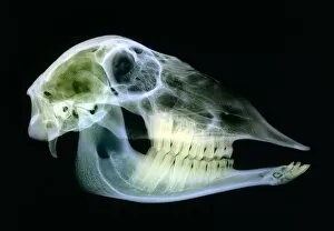
Background imageFalse Coloured Collection: Sheep skull

Sheep skull. Coloured X-ray of a sagittal section through a sheeps (Ovis aries) skull. Sheep have 32 permanent teeth; 12 molars and eight incisors on the bottom jaw and 12 molars on the top jaw

Background imageFalse Coloured Collection: Turtle ants head, SEM

Turtle ants head, SEM
Turtle ants head. Coloured scanning electron micrograph (SEM) of the head of a soldier turtle ant (Cephalotes sp.) from the Amazonian rainforest. One of its compound eyes (green) is at left

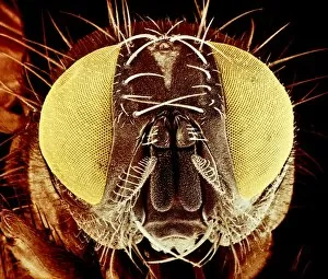
Background imageFalse Coloured Collection: False-colour SEM of the head of a housefly

False-colour SEM of the head of a housefly
Coloured scanning electron micrograph of the head of a female housefly, Musca domestica. The head is dominated by the large compound eyes, here tinted yellow


For sale as Licensed Images

Choose your image, Select your licence and Download the media

"Revealing the Unseen: Exploring the World Through False Colors" Step back in time to medieval alchemy, where a peculiar contraption known as the "Medieval urine wheel" was used to extract hidden secrets from bodily fluids. Fast forward to modern times and witness how science has evolved, unveiling breathtaking wonders like the Orion Nebula. Delve into the microscopic realm, where particle tracks paint a mesmerizing picture of subatomic interactions. Journey across Martian landscapes through the eyes of Spirit rover images, capturing otherworldly vistas that ignite our imagination. Peering into our own genetic blueprint, false colors bring clarity to X and Y chromosomes, unraveling mysteries within our very cells. A horse's skull takes on an ethereal glow as we explore its intricate structure using advanced imaging techniques. Witness synapse nerve junctions come alive with vibrant hues under a transmission electron microscope (TEM), revealing connections that shape our thoughts and actions. A person holding a camera becomes an enigma when captured through X-ray technology – their inner world exposed for all to see. Marvel at tardigrades' resilience as they navigate their microscopic universe under scanning electron microscopy (SEM). Calcareous phytoplankton dance with color in intricate patterns while nerve cells reveal their complexity in stunning detail. Travel back in time to 19th-century Cornwall and discover the harsh reality of tin mining brought vividly to life through false colors – reminding us of humanity's enduring spirit even amidst adversity. In this captivating journey through various realms – from ancient alchemy to distant galaxies, minuscule particles to majestic landscapes – false colors unlock hidden dimensions beyond what meets the eye, inviting us all into a world brimming with wonder and discovery.