
Fibular Collection
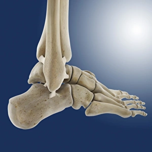
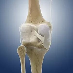
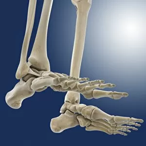
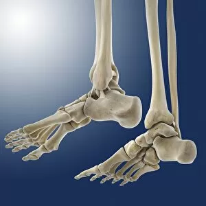
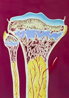
Exploring the intricacies of the human body, let's delve into the world of the fibula - a slim bone in the lower leg

For sale as Licensed Images
Choose your image, Select your licence and Download the media
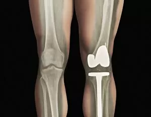
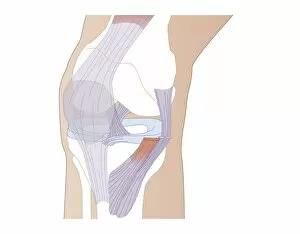
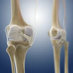
Exploring the intricacies of the human body, let's delve into the world of the fibula - a slim bone in the lower leg. The fibular ligaments, depicted in artwork C013 / 4452 and C013 / 4662, provide stability to the outer ankle. Radiologists meticulously examine knee MRI scans C014 / 1300 to assess any damage to these ligaments, ensuring accurate diagnosis. Total knee replacement X-rays reveal the fibula's role in supporting the replaced joint. Artwork C013 / 4455 and C013 / 4456 provide a closer look at the outer ankle ligaments. Meanwhile, artwork showcasing rickets in the tibia and fibula bones, C013 / 4456, serves as a reminder of the importance of maintaining bone health.