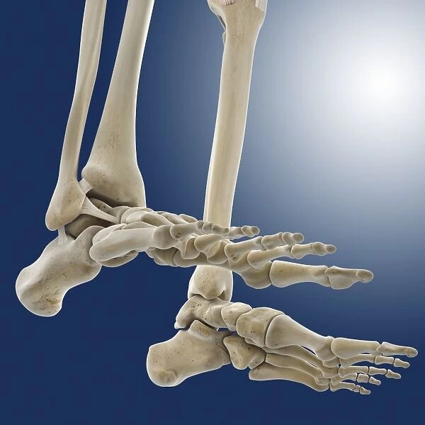
Outer ankle ligaments, artwork C013 / 4456
![]()

Wall Art and Photo Gifts from Science Photo Library
Outer ankle ligaments, artwork C013 / 4456
Outer ankle ligaments. Computer artwork of the bones and ligaments (white) of the feet and ankles from an oblique side view, with the outer side of the right foot at left. The two lower leg bones are the fibula and tibia (to right of fibula). In the foot are the heel bone (calcaneus), the tarsal bones, the metatarsals, and the phalanges (toes). The tibia and fibula articulate with the talus bone in the foot to form the ankle joint. Ligaments are tough bands of fibrous connective tissue that hold the bones of a joint together. These ligaments, named for the bones they join, are the tibiofibular, talofibular, and calcaneofibular
Science Photo Library features Science and Medical images including photos and illustrations
Media ID 9196111
© SPRINGER MEDIZIN/SCIENCE PHOTO LIBRARY
Angular Ankle Arthrology Articulating Articulation Calcaneus Connective Tissue Fibula Fibular Foot Front Frontal Heel Bone Joint Joints Lateral Ligament Ligaments Metatarsal Bones Metatarsals Metatarsus Muscular System Musculoskeletal System Oblique Osteology Phalanges Phalanx Profile Shinbone Tarsals Tibia Toes Navicular Bone
EDITORS COMMENTS
This print showcases the intricate network of outer ankle ligaments in exquisite detail. The computer artwork presents a mesmerizing oblique side view, with the outer side of the right foot elegantly displayed on the left. The fibula and tibia, our lower leg bones, take center stage alongside various other crucial components. From the heel bone (calcaneus) to the metatarsals and phalanges (toes), every element is meticulously depicted for a comprehensive understanding of this vital anatomical region. Ligaments, those resilient bands of fibrous connective tissue responsible for joint stability, are prominently featured throughout this image. Named after the bones they unite, we can observe three significant ligaments: tibiofibular, talofibular, and calcaneofibular. The complexity and sophistication of our musculoskeletal system come to life through this stunning illustration. It serves as a reminder that our bodies are marvels of engineering capable of seamless articulation and movement. Whether you're an anatomy enthusiast or simply intrigued by human biology, this print offers an invaluable glimpse into one aspect of our remarkable physical structure. Appreciate the beauty and intricacy that lies beneath your skin with this extraordinary visual representation from Springer Medizin/Science Photo Library.
MADE IN THE USA
Safe Shipping with 30 Day Money Back Guarantee
FREE PERSONALISATION*
We are proud to offer a range of customisation features including Personalised Captions, Color Filters and Picture Zoom Tools
SECURE PAYMENTS
We happily accept a wide range of payment options so you can pay for the things you need in the way that is most convenient for you
* Options may vary by product and licensing agreement. Zoomed Pictures can be adjusted in the Cart.

