
Glands Collection (#2)
"Glands: Nature's Hidden Gems Unveiled" Botanik Digitalis purpurea L. Fingerhut 160: 1 - Exploring the intricate world of plant glands


For sale as Licensed Images
Choose your image, Select your licence and Download the media
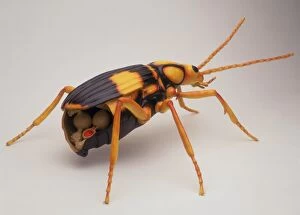
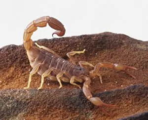
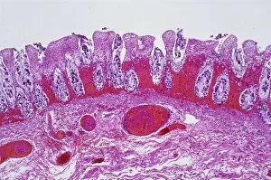
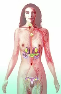
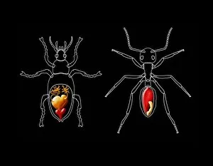
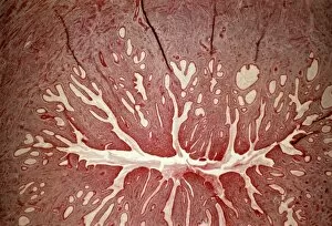
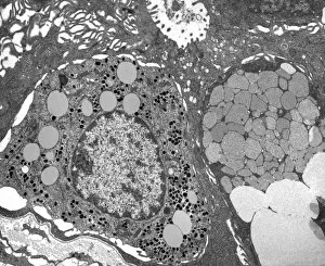
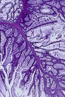
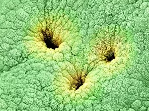
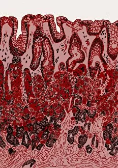
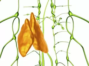
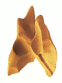
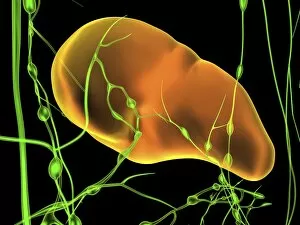
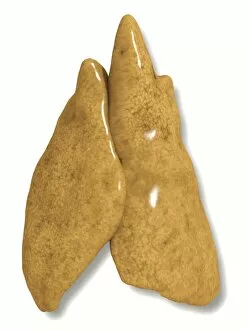
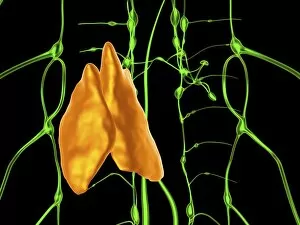
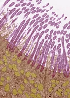
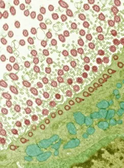
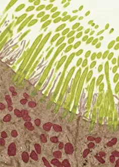
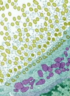
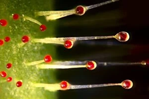
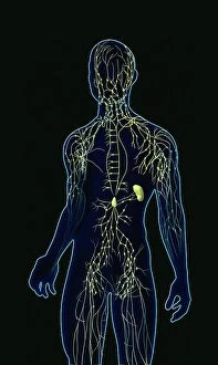
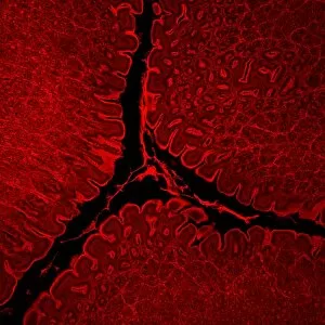
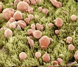
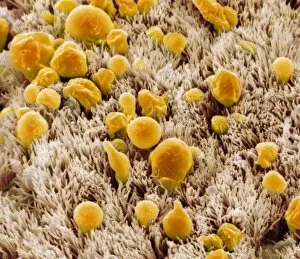
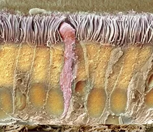
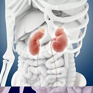
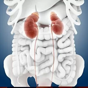
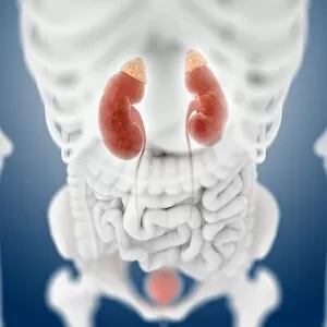
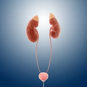
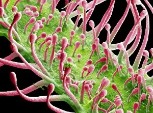
"Glands: Nature's Hidden Gems Unveiled" Botanik Digitalis purpurea L. Fingerhut 160: 1 - Exploring the intricate world of plant glands. French lavender leaf surface, SEM - A closer look at the fascinating structures on a lavender leaf. Leaf oil glands, SEM - Unlocking the secrets behind essential oils through microscopic examination. Orange fruit, light micrograph - Delving into the hidden wonders of an orange's glandular system. Dog anatomy, artwork - Discovering how glands play a vital role in our furry friends' physiology. French lavender leaf surface, SEM (repeated) - Peering into the mesmerizing patterns on a lavender leaf once more. Colony of Bacterium mallei, 1906 (litho) - Examining bacterial colonies and their impact on glandular health. DDE-90036392 – Unraveling the mysteries surrounding specific gland-related research or findings. Frontal View of the Muscles and Glands of Human Neck, 1906 (engraving) – Appreciating our complex neck anatomy and its glandular intricacies throughout history. Girl skipping rope at the beach in white dress – Amidst all scientific exploration lies simple joys like skipping ropes; reminding us that even within ourselves lie remarkable glands waiting to be discovered. From plants to animals and humans alike, these captivating glimpses into various aspects of "glands" shed light on their significance in nature's grand design while reminding us that beauty can be found even in life's tiniest details.