Glands Collection (#3)

"Glands: Nature's Hidden Gems Unveiled" Botanik Digitalis purpurea L. Fingerhut 160: 1 - Exploring the intricate world of plant glands

Background imageGlands Collection: Urinary system, artwork C015 / 6455

Urinary system, artwork C015 / 6455
Urinary system. Computer artwork showing the organs of the human urinary system. The kidneys (red, top left and right) filter waste products such as urea from the blood (red and blue) to form urine

Background imageGlands Collection: Urinary system, artwork C015 / 6454

Urinary system, artwork C015 / 6454
Urinary system. Computer artwork showing the organs of the human urinary system. The kidneys (red, top left and right) filter waste products such as urea from the blood to form urine

Background imageGlands Collection: Tuberculosis, light micrograph C015 / 6400

Tuberculosis, light micrograph C015 / 6400
Tuberculosis. Light micrograph of a section through a lymph node showing a Langhans cell (centre) caused by tuberculosis (TB)

Background imageGlands Collection: Stomach wall, confocal light micrograph C016 / 3127

Stomach wall, confocal light micrograph C016 / 3127
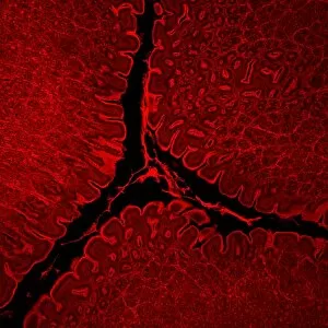
Stomach wall. Laser scanning confocal light micrograph of a section through the wall of a stomach

Background imageGlands Collection: Paediatric examination C013 / 8824

Paediatric examination C013 / 8824
Paediatric examination. Doctor using a stethoscope to listen to a 3-year-old boys lungs. A stethoscope is used to listen to sounds within the body

Background imageGlands Collection: Trachea lining, SEM C013 / 7127

Trachea lining, SEM C013 / 7127
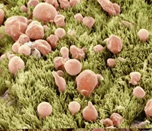
Trachea lining. Coloured scanning electron micrograph (SEM) of the lining of a trachea (wind pipe), showing the cilia (hair-like) and goblet cells (pink). The trachea links the larynx to the lungs

Background imageGlands Collection: Trachea lining, SEM C013 / 7125

Trachea lining, SEM C013 / 7125
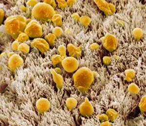
Trachea lining. Coloured scanning electron micrograph (SEM) of the lining of a trachea (wind pipe), showing the cilia (hair-like) and goblet cells (orange). The trachea links the larynx to the lungs

Background imageGlands Collection: Trachea lining, SEM C013 / 7124

Trachea lining, SEM C013 / 7124
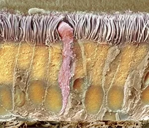
Trachea lining. Coloured scanning electron micrograph (SEM) of a section through the wall of a trachea (wind pipe). The trachea links the larynx to the lungs

Background imageGlands Collection: Trachea lining, SEM C013 / 7123

Trachea lining, SEM C013 / 7123
Trachea lining. Coloured scanning electron micrograph (SEM) of a section through the wall of a trachea (wind pipe). The trachea links the larynx to the lungs

Background imageGlands Collection: Kidneys and ureters, artwork C013 / 4672

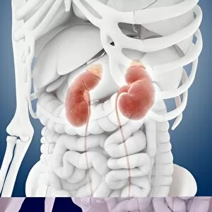
Kidneys and ureters, artwork C013 / 4672
Kidneys and ureters. Computer artwork showing an oblique anterior view of the kidneys, the main organs of the urinary system, relative to the skeleton and abdominal organs (white)

Background imageGlands Collection: Kidneys and ureters, artwork C013 / 4671

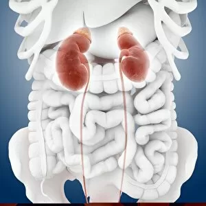
Kidneys and ureters, artwork C013 / 4671
Kidneys and ureters. Computer artwork showing an anterior view of the kidneys, the main organs of the urinary system, relative to the skeleton and abdominal organs (white)

Background imageGlands Collection: Urinary system, artwork C013 / 4673

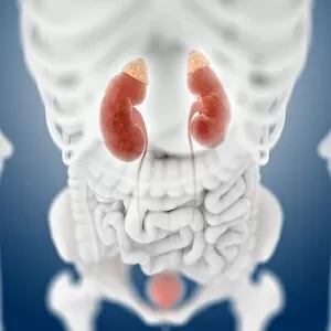
Urinary system, artwork C013 / 4673
Urinary system. Computer artwork showing an anterior view of the kidneys, the main organs of the urinary system, relative to the skeleton and abdominal organs (white)

Background imageGlands Collection: Urinary system, artwork C013 / 4670

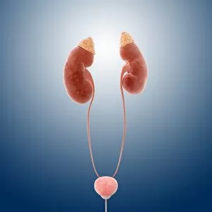
Urinary system, artwork C013 / 4670
Urinary system. Computer artwork showing the kidneys (top), the main organs of the urinary system. The kidneys are located in the upper abdomen

Background imageGlands Collection: Urinary system, artwork C013 / 4669

Urinary system, artwork C013 / 4669
Urinary system. Computer artwork showing the kidneys (top), the main organs of the urinary system. The kidneys are located in the upper abdomen

Background imageGlands Collection: Kidneys and ureters, artwork C013 / 4668

Kidneys and ureters, artwork C013 / 4668
Kidneys and ureters. Computer artwork showing the kidneys, the main organs of the urinary system. The kidneys are located in the upper abdomen

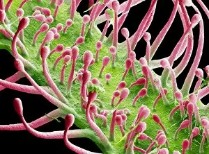
Background imageGlands Collection: Venus flytrap digestive glands, SEM

Venus flytrap digestive glands, SEM
Venus flytrap digestive glands. Coloured scanning electron micrograph (SEM) of digestive glands from a Venus flytrap plant (Dionaea muscipula)

Background imageGlands Collection: Sundew carnivorous plant leaf, SEM

Sundew carnivorous plant leaf, SEM
Sundew leaf. Coloured scanning electron micrograph (SEM) of the sticky stalks (red) on the leaf of a cape sundew (Drosera capensis), a carnivorous plant

Background imageGlands Collection: Perennial Sow-thistle - against the light, showing abundant yellow glandular hairs. Dorset

Perennial Sow-thistle - against the light, showing abundant yellow glandular hairs. Dorset
ROG-14101 Perennial Sow-thistle - against the light, showing abundant yellow glandular hairs. Dorset. UK Sonchus arvensis Bob Gibbons contact details: prints@ardea.com tel: +44 (0) 20 8318 1401

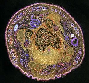
Background imageGlands Collection: Mouse tail, light micrograph

Mouse tail, light micrograph
Mouse tail. Light micrograph of a cross-section through a mouses tail. At centre is the tail vein, which contains red blood cells. A smaller vein (purple squiggle) is seen at top centre left

Background imageGlands Collection: Kidneys and adrenal glands

Kidneys and adrenal glands
Kidneys. Computer artwork of the human renal system. The kidneys (brown/red, left and right) filter waste products and water from blood that passes through the organs in the renal arteries (red)

Background imageGlands Collection: Trachea lining, TEM

Trachea lining, TEM
Trachea lining. Coloured transmission electron micrograph (TEM) of a transverse section through the lining of the trachea (windpipe). The trachea links the larynx to the lungs

Background imageGlands Collection: Brunners glands in the duodenum

Brunners glands in the duodenum
Brunners glands. Fluorescence confocal light micrograph of a section through a human duodenum (small intestine) showing Brunners glands (blue)

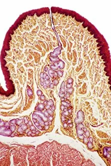
Background imageGlands Collection: Oesophagus wall, light micrograph

Oesophagus wall, light micrograph
Oesophagus wall. Coloured light micrograph of a section through the human oesophagus, which passes food from the mouth to the stomach

Background imageGlands Collection: Colon lining

Colon lining. Fluorescence confocal light micrograph of the lining of a mouse colon (large intestine). The colon starts at the small intestine and ends at the rectum

Background imageGlands Collection: Nose mucosa, light micrograph

Nose mucosa, light micrograph
Nose mucosa. Light micrograph of a section through the nasal mucosa (the tissue that lines the airways of the nose), in the region lying over the nasal concha

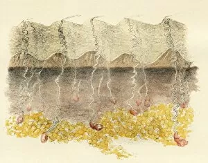
Background imageGlands Collection: Palm

Palm. Magnified section of palm, historical artwork. The external ridged surface is the cuticle, with the thin layer of skin below this known as the rete mucosum, or epidermis

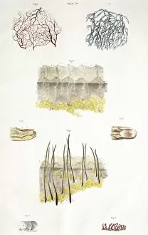
Background imageGlands Collection: Skin anatomy

Skin anatomy. Historical anatomical artwork of the human skin. The main diagram (lower centre) shows scalp hairs and sebaceous glands (yellow, near surface)

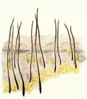
Background imageGlands Collection: Magnified section of scalp

Magnified section of scalp, historical artwork. The external surface is the cuticle, with the layer of skin below this known as the rete mucosum, or epidermis

Background imageGlands Collection: Sarcoidosis of the lungs, X-ray

Sarcoidosis of the lungs, X-ray
Sarcoidosis. Frontal coloured chest X-ray of the lungs of a patient with pulmonary sarcoidosis. Affected lung tissue shows up as red patches

Background imageGlands Collection: Breast implant calcification, X-ray

Breast implant calcification, X-ray
Breast implant calcification. Coloured mammogram (breast X-ray) of calcification around the borders of a breast implant (pink) in a womans breast (red) as seen from the side

Background imageGlands Collection: Swollen lymph nodes, artwork

Swollen lymph nodes, artwork
Lymphatic system, computer artwork

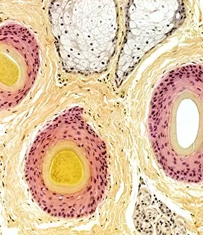
Background imageGlands Collection: Hair follicles, light micrograph

Hair follicles, light micrograph. The hair shaft (orange) is surrounded by an internal root sheath (yellow), an external root sheath (pink) and connective tissue (brown)

Background imageGlands Collection: Human kidneys, artwork

Human kidneys, artwork
Human kidneys, computer artwork

Background imageGlands Collection: Frog skin glands, light micrograph

Frog skin glands, light micrograph
Frog skin glands. Light micrograph of a section through the skin of a frog, showing secretory glands (round, upper centre) just below the surface (across top)

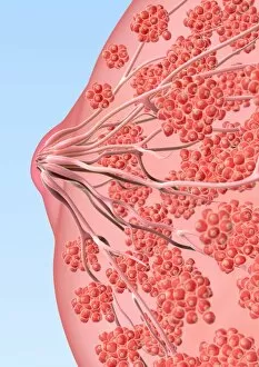
Background imageGlands Collection: Breast tissue, artwork

Breast tissue, artwork
Breast tissue. Computer artwork of a womans breast, showing the mammary glands (red) that produce milk, and the lactiferous ducts (tubes), which carry the milk to the nipple (centre, left)

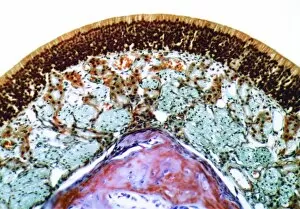
Background imageGlands Collection: Common sundew, light micrograph

Common sundew, light micrograph
Common sundew. Light micrograph of a section through the leaf of a common sundew (Drosera rotundifolia) plant, showing the glandular hairs (dark)


For sale as Licensed Images

Choose your image, Select your licence and Download the media

"Glands: Nature's Hidden Gems Unveiled" Botanik Digitalis purpurea L. Fingerhut 160: 1 - Exploring the intricate world of plant glands. French lavender leaf surface, SEM - A closer look at the fascinating structures on a lavender leaf. Leaf oil glands, SEM - Unlocking the secrets behind essential oils through microscopic examination. Orange fruit, light micrograph - Delving into the hidden wonders of an orange's glandular system. Dog anatomy, artwork - Discovering how glands play a vital role in our furry friends' physiology. French lavender leaf surface, SEM (repeated) - Peering into the mesmerizing patterns on a lavender leaf once more. Colony of Bacterium mallei, 1906 (litho) - Examining bacterial colonies and their impact on glandular health. DDE-90036392 – Unraveling the mysteries surrounding specific gland-related research or findings. Frontal View of the Muscles and Glands of Human Neck, 1906 (engraving) – Appreciating our complex neck anatomy and its glandular intricacies throughout history. Girl skipping rope at the beach in white dress – Amidst all scientific exploration lies simple joys like skipping ropes; reminding us that even within ourselves lie remarkable glands waiting to be discovered. From plants to animals and humans alike, these captivating glimpses into various aspects of "glands" shed light on their significance in nature's grand design while reminding us that beauty can be found even in life's tiniest details.