Golgi Apparatus Collection (#2)

The Golgi apparatus, a vital organelle in animal and plant cells, plays a crucial role in the processing and packaging of proteins

Background imageGolgi Apparatus Collection: Animal cell organelles, artwork C016 / 0612

Animal cell organelles, artwork C016 / 0612
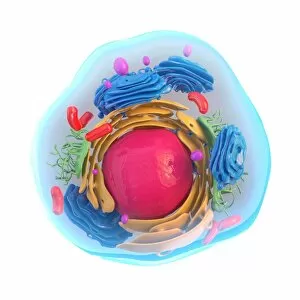
Computer artwork showing the organelles in a eukaryotic cell. This is an animal cell. Structures include the nucleus (centre, orange) which has a membrane with nuclear pores

Background imageGolgi Apparatus Collection: Animal cell organelles, artwork C016 / 0605

Animal cell organelles, artwork C016 / 0605
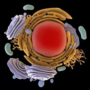
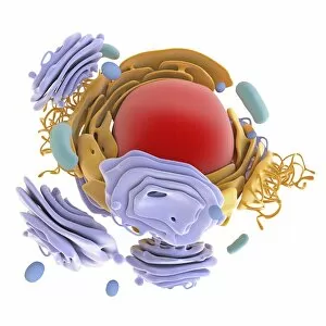
Computer artwork showing the organelles in a eukaryotic cell. This is an animal cell. Structures include the nucleus (centre, red) which has a membrane with nuclear pores

Background imageGolgi Apparatus Collection: Animal cell organelles, artwork C016 / 0610

Animal cell organelles, artwork C016 / 0610
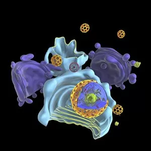
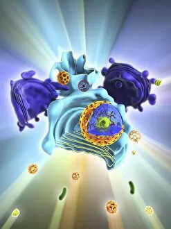
Computer artwork showing the organelles in a eukaryotic cell. This is an animal cell. Structures include the nucleus (centre, orange) which has a membrane with nuclear pores

Background imageGolgi Apparatus Collection: Animal cell organelles, artwork C016 / 0604

Animal cell organelles, artwork C016 / 0604
Computer artwork showing the organelles in a eukaryotic cell. This is an animal cell. Structures include the nucleus (centre, red) which has a membrane with nuclear pores

Background imageGolgi Apparatus Collection: Animal cell organelles, artwork C016 / 0606

Animal cell organelles, artwork C016 / 0606
Computer artwork showing the organelles in a eukaryotic cell. This is an animal cell. Structures include the nucleus (centre, orange) which has a membrane with nuclear pores

Background imageGolgi Apparatus Collection: Animal cell, artwork C013 / 9985

Animal cell, artwork C013 / 9985
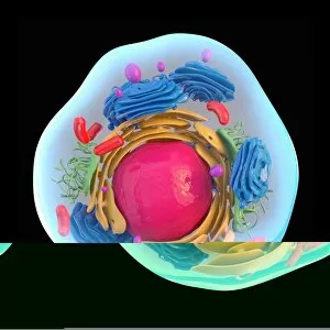
Animal cell. Computer artwork showing the cell organelles found inside a typical animal cell. The nucleus (large round) can be seen at centre

Background imageGolgi Apparatus Collection: Animal cell, artwork C013 / 9984

Animal cell, artwork C013 / 9984
Animal cell. Computer artwork showing the cell organelles found inside a typical animal cell. The nucleus (large round) can be seen at centre

Background imageGolgi Apparatus Collection: Human cell, artwork C013 / 6522

Human cell, artwork C013 / 6522
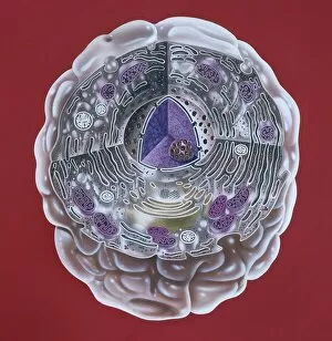
Human cell. Illustration of a section through a human cell. At centre is the nucleus (purple), with the nucleolus (dark red) inside it

Background imageGolgi Apparatus Collection: Intracellular transport, artwork C013 / 4997

Intracellular transport, artwork C013 / 4997
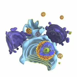
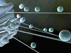
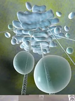
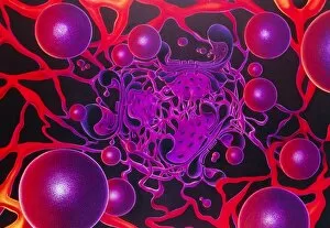
Intracellular transport. Computer artwork of vesicles (spheres) being transported from a Golgi body (blue, left) around the cell by microtubules (string-like)

Background imageGolgi Apparatus Collection: Intracellular transport, artwork C013 / 4995

Intracellular transport, artwork C013 / 4995
Intracellular transport. Computer artwork of vesicles (spheres) being transported from a Golgi body (blue, top) around the cell by microtubules (string-like)

Background imageGolgi Apparatus Collection: Brain cells, TEM C013 / 4801

Brain cells, TEM C013 / 4801
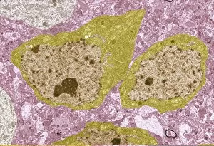
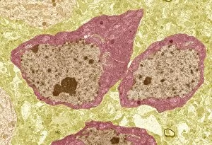
Brain cells. Transmission electron micrograph (TEM) of a section through oligodendrocytes (dark) in human brain tissue, showing free ribosomes (dark green dots), golgi apparatus (curved brown lines)

Background imageGolgi Apparatus Collection: Brain cells, TEM C013 / 4800

Brain cells, TEM C013 / 4800
Brain cells. Transmission electron micrograph (TEM) of a section through oligodendrocytes in human brain tissue, showing free ribosomes (dark pink dots), golgi apparatus (curved brown lines)

Background imageGolgi Apparatus Collection: Brain cell, TEM C013 / 4799

Brain cell, TEM C013 / 4799
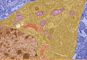
Brain cell. Transmission electron micrograph (TEM) of a section through an oligodendrocyte in human brain tissue, showing free ribosomes (dark brown dots), golgi apparatus (curved orange lines)

Background imageGolgi Apparatus Collection: Brain cell, TEM C013 / 4798

Brain cell, TEM C013 / 4798
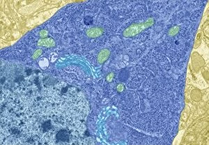
Brain cell. Transmission electron micrograph (TEM) of a section through an oligodendrocyte in human brain tissue, showing free ribosomes (dark blue dots), golgi apparatus (curved light blue lines)

Background imageGolgi Apparatus Collection: Artwork of a human egg cell

Artwork of a human egg cell in the primordial follicle phase which occurs after seven months of fetal development. The egg cell is surrounded by a thick layer of follicular cells known as zona

Background imageGolgi Apparatus Collection: Cell mitosis

Cell mitosis. Confocal fluorescence light micrograph composite showing 6 stages of mitotic cell division. At far left, the cell has completed the first stage of cell division known as interphase in

Background imageGolgi Apparatus Collection: Pancreas cell, TEM

Pancreas cell, TEM
Pancreas cell. Coloured transmission electron micrograph (TEM) of an acinar (exocrine) pancreatic cell. Acinar cells secrete the inactive precursors (zymogens)

Background imageGolgi Apparatus Collection: Golgi apparatus, TEM

Golgi apparatus, TEM
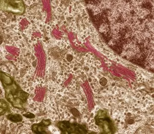
Golgi apparatus. Coloured transmission electron micrograph (TEM) of Golgi apparatus, also known as Golgi bodies, (pink) in a macrophage white blood cell

Background imageGolgi Apparatus Collection: Inside of an imaginary cell

Inside of an imaginary cell
Inside a cell. Panoramic view from inside an imaginary cell. The densely folded endoplasmic reticulum, a membrane that divides the cell into compartments, is shown in yellow and red

Background imageGolgi Apparatus Collection: Animal cell structure, computer artwork

Animal cell structure, computer artwork
Animal cell. Computer artwork of a section through an animal cell. At the centre is the nucleus, which contains the cells genetic information in the form of DNA (deoxyribonucleic acid)

Background imageGolgi Apparatus Collection: Artwork of ER, golgi body, plasma membrane of cell

Artwork of ER, golgi body, plasma membrane of cell
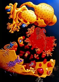
Illustration of membrane structures in a cell: endoplasmic reticulum, golgi apparatus and plasma membrane. At top, the nucleus is seen with pores, & ribosomes (pink dots) on its surface

Background imageGolgi Apparatus Collection: Cell organelles, TEM

Cell organelles, TEM
Cell organelles. Coloured transmission electron micrograph (TEM) of several organelles in an animal cell. At centre, the nucleus (dark purple, round) contains the cells genetic material (DNA)

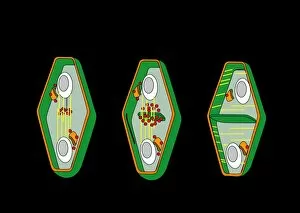
Background imageGolgi Apparatus Collection: Cytokinesis, artwork

Cytokinesis, artwork
Cytokinesis. Artwork showing the stage of cell division that involves the splitting of the cell cytoplasm between two daughter cells

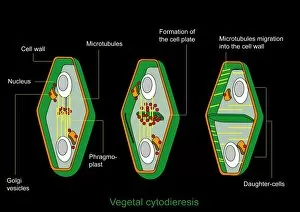
Background imageGolgi Apparatus Collection: Cytokinesis, diagram

Cytokinesis, diagram
Cytokinesis. Diagram showing the stage of cell division that involves the splitting of the cell cytoplasm between two daughter cells

Background imageGolgi Apparatus Collection: Nerve cell anatomy, artwork

Nerve cell anatomy, artwork
Nerve cell anatomy. Computer artwork showing the organelles in a neurone (nerve cell). At centre is nucleus (grey) and nucleolus (black)

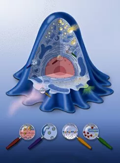
Background imageGolgi Apparatus Collection: Animal cell processes, artwork

Animal cell processes, artwork
Animal cell processes. Cutaway artwork showing the structures inside an animal cell and four different processes that take place inside it or on its membrane (all marked by magnifying glasses)

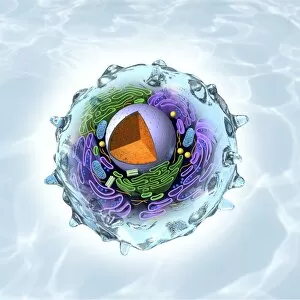
Background imageGolgi Apparatus Collection: Animal cell structure, artwork

Animal cell structure, artwork
Animal cell structure. Artwork showing the internal structure of an animal cell. Inside the cell, the cellular structures (organelles) include the Golgi apparatus (green)

Background imageGolgi Apparatus Collection: Golgi apparatus, artwork

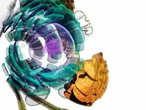
Golgi apparatus, artwork. This structure is an organelle found within eukaryotic cells. It receives proteins and lipids that are synthesised elsewhere on the endoplasmic reticulum

Background imageGolgi Apparatus Collection: Retinal rod cell anatomy, diagram

Retinal rod cell anatomy, diagram

Background imageGolgi Apparatus Collection: Milk-producing cell, artwork

Milk-producing cell, artwork
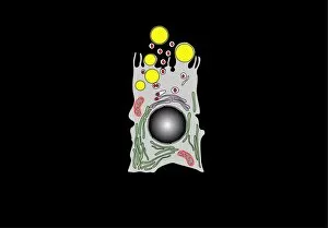
Milk-producing cell. Artwork of the anatomical structure of a lacteal (milk-producing) cell. Normal cell organelles include the nucleus (black, centre), the golgi apparatus (white)

Background imageGolgi Apparatus Collection: Retinal rod cell anatomy, artwork

Retinal rod cell anatomy, artwork
Retinal rod cell anatomy. Artwork of the external (left) and internal (right) anatomical structure of a retinal cell. These cells are found in the retina that lines the back of the eyes

Background imageGolgi Apparatus Collection: Milk-producing cell, diagram

Milk-producing cell, diagram
Milk-producing cell. Diagram of the anatomical structure of a lacteal (milk-producing) cell. Normal cell organelles include the nucleus (black, centre), the golgi apparatus (white)

Background imageGolgi Apparatus Collection: Animal cell, artwork

Animal cell, artwork
Animal cell. Computer artwork of an expanded animal cell. At centre left is the nucleus (purple sphere), which contains the cells genetic information in the form of DNA (deoxyribonucleic acid)

Background imageGolgi Apparatus Collection: Apoptosis, fluorescent light micrograph

Apoptosis, fluorescent light micrograph
Apoptosis. 2-photon fluorescence light micrograph of a cultured cell (centre) undergoing apoptosis, or programmed cell death. It is surrounded by healthy cells


For sale as Licensed Images

Choose your image, Select your licence and Download the media

The Golgi apparatus, a vital organelle in animal and plant cells, plays a crucial role in the processing and packaging of proteins. This complex structure is intricately connected to other cellular components like the rough endoplasmic reticulum (ER), as shown in transmission electron microscopy (TEM) images. In HeLa cells, captured through light micrograph C017 / 8298, we can observe the Golgi apparatus's distinctive appearance. Interestingly, this organelle is not limited to animal cells alone; it also exists in trypanosome protozoans as depicted in stunning artwork. Similarly, scanning electron microscopy (SEM) allows us to visualize the intricate details of the Golgi apparatus within fibroblast cells through captivating artwork. Understanding animal cell structures is essential for comprehending their functions fully. Hepatocyte liver cells imaged using TEM provide valuable insights into how the Golgi apparatus operates within these specialized cells. Cross-sectional illustrations of human cells further enhance our understanding of this remarkable organelle's location and function. In plant cells, which possess unique features compared to animal cells, the Golgi apparatus coexists with other essential components such as nuclei, nucleoli, ribosomes, and endoplasmic reticulum. These elements are beautifully illustrated in conceptual images that showcase both plant cell structures and their individual components. Whether it be an artistic representation or a conceptual image capturing its essence within different organisms' cellular contexts—be it animals or plants—the Golgi apparatus remains an intriguing subject for scientific exploration. Its significance cannot be overstated when considering protein synthesis and secretion pathways critical for various biological processes across diverse species.