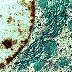
Brain cell, TEM C013 / 4799

![]()
Wall Art and Photo Gifts from Science Photo Library
Brain cell, TEM C013 / 4799
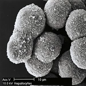
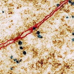
Brain cell. Transmission electron micrograph (TEM) of a section through an oligodendrocyte in human brain tissue, showing free ribosomes (dark brown dots), golgi apparatus (curved orange lines), lysosomes (brown circles) and mitochondria (purple). The cell nucleus (large, dark brown) is a lower left. Oligodendrocytes occur in both the white and grey matter of the central nervous system (CNS). Their main function is to provide support and to form myelin sheaths around neurons (nerve cells), which insulates the axon of each cell, allowing efficient transmission of electrical impulses. Magnification: x20, 000 at 10 centimetres wide
Science Photo Library features Science and Medical images including photos and illustrations
Media ID 9195019
© STEVE GSCHMEISSNER/SCIENCE PHOTO LIBRARY
Cell Biology Central Nervous System Cytological Cytology Endoplasmic Reticulum Glia Glial Golgi Apparatus Histological Histology Insulating Insulation Lysosome Lysosomes Microglia Microglial Mitochondria Mitochondrion Myelin Sheath Myelinated Myelination Nerve Cell Neuroglia Nuclei Nucleus Oligodendrocyte Ribosome Ribosomes Tissue Transmission Electron Micrograph Transmission Electron Microscope Brain Nervous System Neurological Neurology
EDITORS COMMENTS
This photo print, titled "Brain cell, TEM C013 / 4799" offers a mesmerizing glimpse into the intricate world of human brain tissue. Taken using a transmission electron microscope (TEM), it showcases a section through an oligodendrocyte, one of the essential cells found in both white and grey matter of the central nervous system (CNS). The image reveals various cellular components with remarkable clarity. Dark brown dots represent free ribosomes, while curved orange lines depict the golgi apparatus. Brown circles signify lysosomes, and purple structures symbolize mitochondria. The large, dark brown nucleus rests at the lower left corner. Oligodendrocytes play a crucial role in supporting neurons by forming myelin sheaths around them. These sheaths act as insulation for axons, enabling efficient transmission of electrical impulses throughout the nervous system. With its magnification set at x20,000 and measuring 10 centimeters wide, this photograph provides an awe-inspiring view into the microscopic realm of biology and anatomy. It highlights not only the complexity but also the beauty inherent within our neural architecture. Captured by renowned photographer Steve Gschmeissner from Science Photo Library's collection, this image serves as a testament to humanity's ongoing exploration and understanding of our own intricate biology.
MADE IN THE USA
Safe Shipping with 30 Day Money Back Guarantee
FREE PERSONALISATION*
We are proud to offer a range of customisation features including Personalised Captions, Color Filters and Picture Zoom Tools
SECURE PAYMENTS
We happily accept a wide range of payment options so you can pay for the things you need in the way that is most convenient for you
* Options may vary by product and licensing agreement. Zoomed Pictures can be adjusted in the Cart.