
Granules Collection (#2)
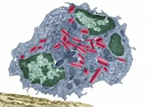
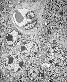
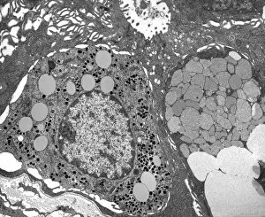
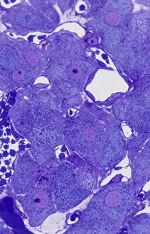
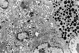
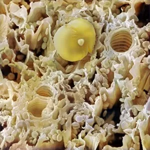
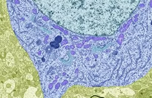
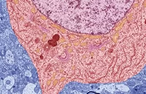
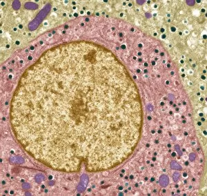
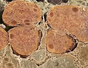
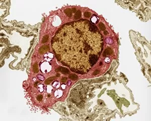
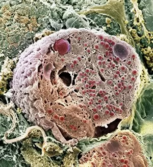
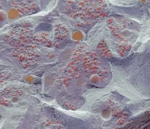
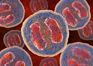
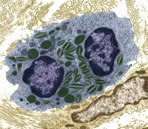
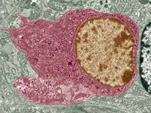
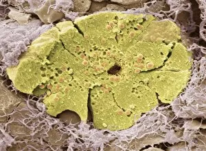
Granules are tiny, granular structures that play a crucial role in various aspects of our lives


For sale as Licensed Images
Choose your image, Select your licence and Download the media
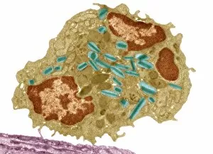
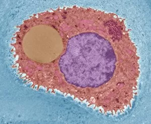
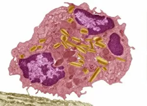
Granules are tiny, granular structures that play a crucial role in various aspects of our lives. In the world of biology, basophil white blood cells contain these granules, which are essential for their function in the immune system. These specialized cells help to defend our bodies against foreign invaders and allergens. When examining a blood cell under a microscope, one might come across intriguing features known as Dohle bodies. These small blue-gray they are be observed within the cytoplasm of neutrophils - another type of white blood cell. Their presence often indicates an ongoing infection or inflammation within the body. Micrographs provide us with detailed images that allow us to explore the intricate world even further. Picture No. 11014616 captures these microscopic structures in all their glory, showcasing their unique shapes and sizes. While focusing on granules is fascinating, it's also important to take breaks from scientific exploration and appreciate nature's beauty. Lighthouse Scenic in Northumberland, England (Picture No. 11014611) offers a breathtaking view where we can escape from laboratory settings and immerse ourselves in serene surroundings. As we continue our journey through captivating visuals, Picture No. 11014610 transports us to picturesque landscapes adorned with vibrant colors and textures – reminding us that even nature possesses its own version of granular formations. Moving forward along this visual adventure (Picture No. s 11014609-11014600), we encounter diverse scenes ranging from tranquil beaches to lush forests – each displaying its own distinct charm while reminding us that life itself is composed of countless tiny fragments coming together harmoniously. Whether exploring microscopic wonders like basophil white blood cells or marveling at stunning natural landscapes like those found in Northumberland, England – there is no denying the significance and beauty held within every grain-like structure we encounter throughout our existence.