Haematology Collection (#9)

Haematology, the captivating study of blood cells and their intricate functions, unveils a world unseen to the naked eye

Background imageHaematology Collection: Blood cells, computer artwork

Blood cells, computer artwork
Blood cells. Computer artwork of red blood cells, white blood, white blood cells (blue) and platelets (yellow) travelling through the lumen of the blood vessel

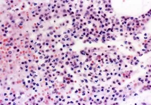
Background imageHaematology Collection: Human blood cells, light micrograph

Human blood cells, light micrograph
Human blood cells. Light micrograph of blood cells at a site of inflammation, showing red blood cells (erythrocytes, red) surrounded by many granulocytes (blue)

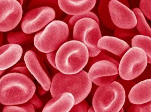
Background imageHaematology Collection: Red blood cells, SEM

Red blood cells, SEM
Red blood cells. Coloured scanning electron micrograph (SEM) of red blood cells (erythrocytes). Red blood cells are biconcave, disc-shaped cells that transport oxygen from the lungs to body cells

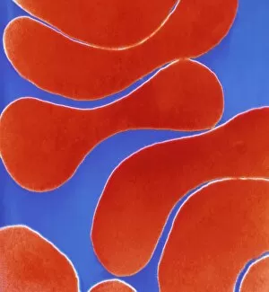
Background imageHaematology Collection: Colour TEM of red blood cells, rouleau formation

Colour TEM of red blood cells, rouleau formation
Red blood cells. Coloured Transmission Electron Micrograph (TEM) of sectioned human red blood cells (erythrocytes) in a rouleau formation

Background imageHaematology Collection: Types of blood cell

Types of blood cell
Blood cells. Artwork (based on a scanning electron micrograph) showing various blood cells escaping from a sectioned small vein (top right)

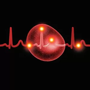
Background imageHaematology Collection: ECG and red blood cells

ECG and red blood cells. Computer artwork of an electrocardiogram (ECG) trace and red blood cells (erythrocytes). Red blood cells are biconcave, giving them a large surface area for gas exchange

Background imageHaematology Collection: ECG and red blood cell

ECG and red blood cell. Computer artwork of an electrocardiogram (ECG) trace and a red blood cell (erythrocyte). Red blood cells are biconcave, giving them a large surface area for gas exchange

Background imageHaematology Collection: Red blood cells and ECG

Red blood cells and ECG. Computer artwork of red blood cells (erythrocytes), an electrocardiogram (ECG) trace and a silhouette of a person

Background imageHaematology Collection: Blood vessel leak, TEM

Blood vessel leak, TEM
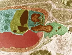
Blood vessel leak. Coloured transmission electron micrograph (TEM) of a white blood cell (centre, green and brown) leaking out of a blood vessel

Background imageHaematology Collection: Artery and red blood cells

Artery and red blood cells. Computer artwork of an artery, showing red blood cells (erythrocytes). These are specialised cells that carry oxygen from the lungs to body tissues

Background imageHaematology Collection: Arteriole, SEM

Arteriole, SEM
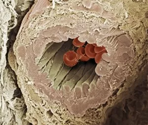
Arteriole. Coloured scanning electron micrograph (SEM) of a cross-section through a small human artery known as an arteriole. Red blood cells and some fibrin fibres are seen in the central lumen

Background imageHaematology Collection: Blood transfusion

Blood transfusion
MODEL RELEASED. Blood transfusion. Blood-filled tubing attached to a patients arm during a blood transfusion. A needle (centre, underneath the dressing) has been placed in a vein

Background imageHaematology Collection: Blood transfusion equipment

Blood transfusion equipment. Bags of A positive blood hanging on a stand and being used in a blood transfusion. Blood transfusions are carried out to replace blood in people with blood disorders

Background imageHaematology Collection: Blood test

Blood test. Drop of blood on a watchglass. Beneath the watchglass is a blood test result sheet

Background imageHaematology Collection: Blood analysis, conceptual artwork

Blood analysis, conceptual artwork
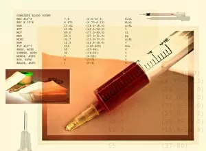
Blood testing, conceptual artwork. A syringe is used to take a sample of blood for testing. At upper left are the levels of some blood cells in the tested blood

Background imageHaematology Collection: Centrifuged blood

Centrifuged blood. Technician holding a vacutainer tube filled with centrifuged blood. A centrifuge is a machine that spins its contents around a central axis at very high speeds

Background imageHaematology Collection: Blood centrifuge preparation

Blood centrifuge preparation
Blood sample being placed in a container before going into a centrifuge. A centrifuge is a machine that spins its contents around a central axis at very high speeds

Background imageHaematology Collection: Blood testing

Blood testing
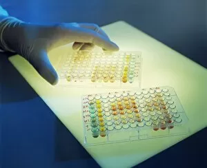
Blood analysis. Gloved hand with multi-well plates containing blood for analysis. Blood can be analysed to check for the presence or absence of antigens, allergens, drugs or pathogenic organisms

Background imageHaematology Collection: AIDS blood testing

AIDS blood testing
MODEL RELEASED. AIDS blood test. Technician holding a multi-well sample tray as he tests blood for HIV. HIV (human immunodeficiency virus) causes AIDS (acquired immune deficiency syndrome)

Background imageHaematology Collection: Deep vein thrombosis, artwork

Deep vein thrombosis, artwork
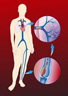
Deep vein thrombosis. Computer artwork showing blood clots in a leg (brown lumps, bottom right), caused by deep vein thrombosis (DVT), moving to blood vessels in the lungs (blue, top right)

Background imageHaematology Collection: Abnormal blood clot, TEM

Abnormal blood clot, TEM
Abnormal blood clot. Coloured transmission electron micrograph (TEM) of a section through a blood vessel that is blocked by an abnormal blood clot (thrombus)

Background imageHaematology Collection: Malaria, conceptual artwork

Malaria, conceptual artwork. Drop of red blood, showing the outline of the continent of Africa. Malaria is widespread in tropical and subtropical regions, including parts of the Americas, Asia

Background imageHaematology Collection: SEM of blood clot

SEM of blood clot
Thrombosis. False-colour scanning electron micrograph (SEM) of a thrombus (clot) of red blood cells inside a blood vessel

Background imageHaematology Collection: Coronary thrombosis, light micrograph

Coronary thrombosis, light micrograph
Coronary thrombosis. Light micrograph of a section through a thrombus (blood clot) in the coronary artery of the heart. The clot is packed full of red blood cells (red)

Background imageHaematology Collection: Anaemia, conceptual artwork

Anaemia, conceptual artwork
Anaemia, conceptual computer artwork. Red blood cell (erythrocyte) with missing iron panels from its surface in front of normal blood cells, representing anaemia

Background imageHaematology Collection: Ebola virus and blood cells

Ebola virus and blood cells. Computer artwork of Ebola viruses particles (yellow) attacking a human white blood cell. Red blood cells are also seen

Background imageHaematology Collection: Activated platelet, SEM

Activated platelet, SEM
Activated platelet. Coloured scanning electron micrograph (SEM) of an activated platelet, or thrombocyte, from peripheral blood. Magnification: x2, 500 when printed at 10 centimetres wide

Background imageHaematology Collection: Taking a blood sample

Taking a blood sample
PROPERTY RELEASED. MODEL RELEASED. Taking a blood sample

Background imageHaematology Collection: Bone marrow stem cell, SEM

Bone marrow stem cell, SEM
Bone marrow stem cell, coloured scanning electron micrograph (SEM). This cell is known as a multipotential stem cell because it can form the precursors to every type of blood cell


For sale as Licensed Images

Choose your image, Select your licence and Download the media

Haematology, the captivating study of blood cells and their intricate functions, unveils a world unseen to the naked eye. Through scanning electron microscopy (SEM), we delve into this mesmerizing realm where red and white blood cells take center stage. Witnessing these tiny heroes in action is truly awe-inspiring. The blood coagulation cascade, an essential process for wound healing, comes alive through artwork C016 / 9873. Each step meticulously depicted, showcasing the complexity behind clot formation. Meanwhile, SEM reveals the delicate beauty of red blood cells as they flow gracefully through our veins. Dohle bodies in a blood cell micrograph offer a glimpse into abnormalities that can occur within these vital components of life itself. Their presence hints at underlying health conditions that require further investigation. Acute promyelocytic leukaemia's impact becomes evident when examining its micrograph closely. The distorted appearance of affected cells serves as a stark reminder of the challenges faced by those battling this disease. Intriguingly, even on plaster surfaces under SEM scrutiny, we uncover unexpected encounters with blood clots - reminders that our body's defense mechanisms are always at work to protect us from harm. Computer artwork brings forth vibrant depictions of red blood cells dancing harmoniously together; their collective effort ensuring oxygen reaches every corner of our being. A testament to nature's remarkable design and efficiency. And let us not forget about stem cells – versatile powerhouses capable of transforming into various specialized cell types. SEM provides an up-close look at these extraordinary entities that hold immense potential for regenerative medicine and groundbreaking therapies. Finally, an enchanting connection between red blood cells and the heart emerges before our eyes – symbolizing life's unbreakable bond between circulation and vitality. Haematology unravels secrets hidden within each drop coursing through our veins - reminding us how intricately woven science is with human existence itself.