
Hepatic Collection
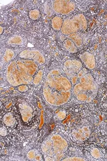
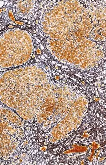
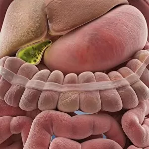
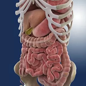
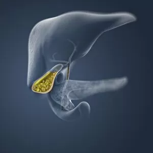
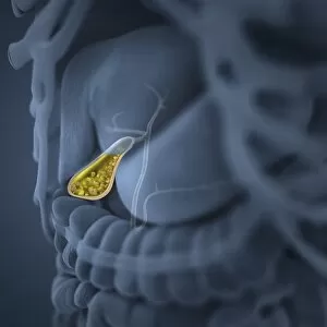
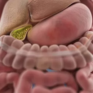
The hepatic system, a vital component of our body's functioning, is often associated with liver cirrhosis


For sale as Licensed Images
Choose your image, Select your licence and Download the media
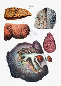
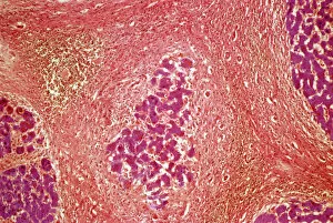
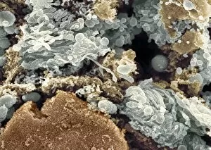
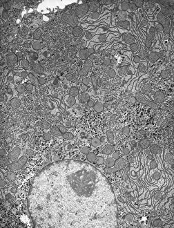
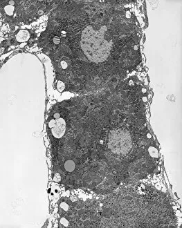
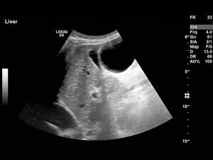
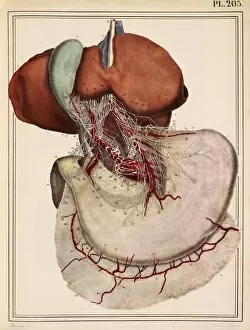
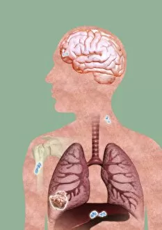
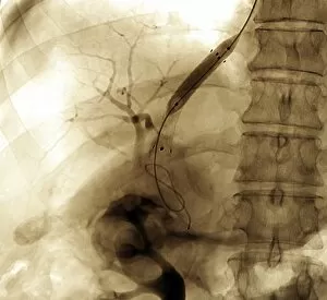
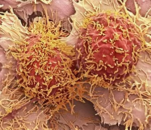
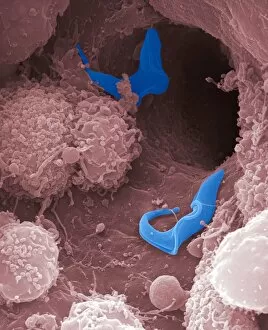
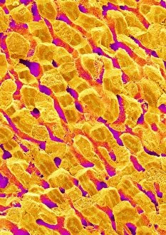
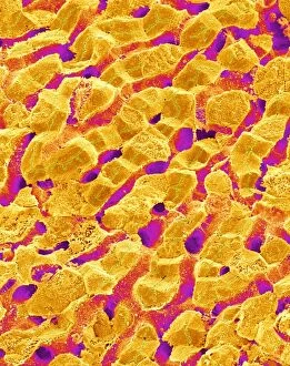
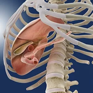
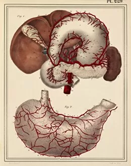
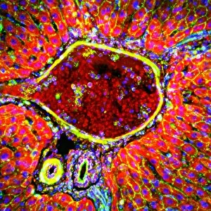
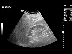
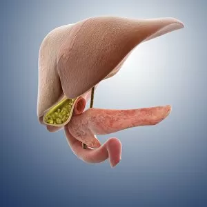
The hepatic system, a vital component of our body's functioning, is often associated with liver cirrhosis. This condition, characterized by the scarring of liver tissue, can be observed under a light microscope as a mesmerizing pattern resembling an abstract painting. Meanwhile, the Golgi apparatus within hepatocytes plays a crucial role in protein synthesis and secretion - its intricate structure captured beautifully through scanning electron microscopy. Intriguing X-ray images reveal the network of abdominal arteries that supply blood to this remarkable organ. The abdomen itself houses various organs working harmoniously to maintain our well-being. Among them stands the Endive Pellia liverwort, gracefully growing amidst Common Liverwort - showcasing nature's resilience and adaptability. On another note, an artistic lithograph showcases the hand's diverse forms of fingers and palmistry lines - reminding us of how unique each individual is. Ultrasound scans provide valuable insights into different conditions affecting the liver: from detecting liver masses to diagnosing polycystic liver disease. Lastly, an 1825 artwork depicts the intricate connection between nerves in both the liver and stomach - emphasizing their interdependence for proper digestion and overall health. Through these captivating glimpses into hepatic aspects ranging from microscopic structures to macroscopic functions, we gain appreciation for this extraordinary organ's complexity and importance in sustaining life.