Immune System Collection (#28)

"The Immune System: A Complex Network of Defenders" Our a remarkable defense mechanism that safeguards our body against harmful invaders

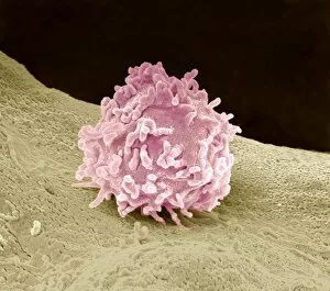
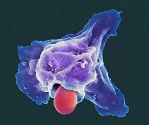
Background imageImmune System Collection: Macrophage cell, SEM

Macrophage cell, SEM
Macrophage cell. Coloured scanning electron micrograph (SEM) of a macrophage white blood cell. Macrophages are cells of the bodys immune system
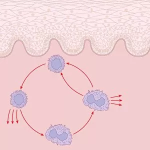
Background imageImmune System Collection: Immune response to chronic inflammation

Immune response to chronic inflammation. Computer artwork showing the interaction of T cell, lymphocyte, and macrophage white blood cells in response to chronic inflammation of the skin (top)

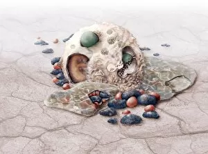
Background imageImmune System Collection: Skin damage immune response, artwork

Skin damage immune response, artwork
Skin damage immune response. Computer artwork showing the bodys immune response to pathogens (red dots) entering through a skin wound (upper centre)

Background imageImmune System Collection: Neutrophil engulfing fungus, SEM

Neutrophil engulfing fungus, SEM
Neutrophil engulfing fungus. Coloured scanning electron micrograph (SEM) of a neutrophil white blood cell (orange) engulfing and destroying a Candida albicans (thrush) fungal hypha (green)

Background imageImmune System Collection: Liver macrophage cell, SEM

Liver macrophage cell, SEM
Liver macrophage cell. Coloured scanning electron micrograph (SEM) of a liver macrophage, also known as a Kupffer cell. Macrophages are a type of large white blood cell that recognise foreign

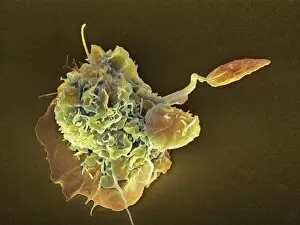
Background imageImmune System Collection: Macrophage attacking a foreign body, SEM

Macrophage attacking a foreign body, SEM
Macrophage attacking a foreign body. Coloured scanning electron micrograph (SEM) of a macrophage white blood cell (round, left) engulfing a Leishmania mexicana protozoan parasite (long, right)

Background imageImmune System Collection: Granulocyte, SEM

Granulocyte, SEM
Granulocyte. Coloured scanning electron micrograph (SEM) of a granulocyte in bone marrow. Granulocytes are a category of white blood cell characterized by the presence of granules in their cytoplasm

Background imageImmune System Collection: Granulocytes, SEM

Granulocytes, SEM
Granulocytes. Coloured scanning electron micrograph (SEM) of granulocytes in bone marrow. Granulocytes are a category of white blood cell characterized by the presence of granules in their cytoplasm

Background imageImmune System Collection: Woundcare gel therapy, artwork

Woundcare gel therapy, artwork
Woundcare gel therapy. Artwork showing the cellular effects of woundcare therapy using a platelet-leucocyte enriched gel to support soft tissue and bone healing

Background imageImmune System Collection: Parasite and cells

Parasite and cells
Concept image. An aggresive unknown Parasite has entered a cell and consumes it from within

Background imageImmune System Collection: Thoracic lymphatic system, artwork

Thoracic lymphatic system, artwork
Thoracic lymphatic system. Artwork of a profile view of the thorax showing the lymphatic system and lymph nodes (red and white ovals). The lymphatic system is part of the immune system

Background imageImmune System Collection: Allergic immune response, artwork

Allergic immune response, artwork
mast cell reponse: Bacteria (blue) bounds to a mast cell (White ball) and histamine (orange) was triggered

Background imageImmune System Collection: Interferon regulatory factor molecule

Interferon regulatory factor molecule
Interferon regulatory factor. Molecular model of interferon regulatory factor 3 (IRF3, blue and white) bound to a DNA (deoxyribonucleic acid) molecule (yellow)

Background imageImmune System Collection: Allergic response, artwork

Allergic response, artwork
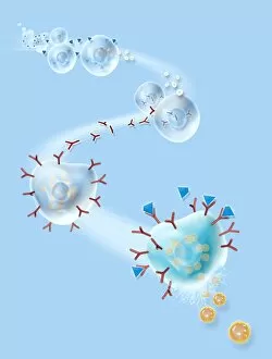
Allergic response. Computer artwork showing the process that produces an allergic reaction. Antigens (blue), such as pollen

Background imageImmune System Collection: Antibodies and their antigen

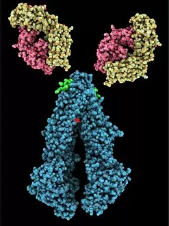
Antibodies and their antigen. Computer model showing the molecular structure of two anti-P-glycoprotein antibodies (upper left and right) and the P-glycoprotein (blue) to which they respond

Background imageImmune System Collection: Elie Metchnikoff, Russian biologist

Elie Metchnikoff, Russian biologist
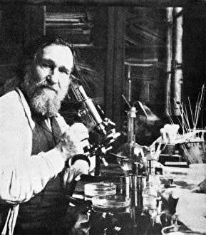
Elie Metchnikoff (1845-1916), Russian biologist and Nobel laureate. Metchnikoff, also known as Ilya Ilyich Mechnikov, is best known for his pioneering research into the immune system

Background imageImmune System Collection: Interferon gamma molecule and receptor

Interferon gamma molecule and receptor
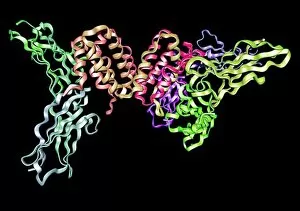
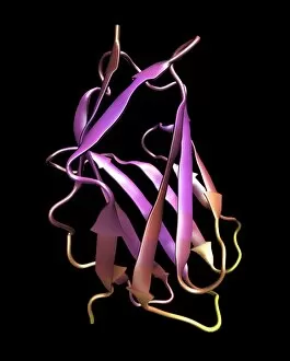
Interferon gamma. Molecular model showing the secondary structure of the interferon gamma dimer (bright pink and gold and pale pink and beige)

Background imageImmune System Collection: Interferon antagonism by viral protein

Interferon antagonism by viral protein
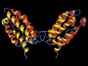
Interferon (IFN) antagonism by viral protein. Molecular model showing two views of an orthopoxvirus IFN-gamma-binding protein bound to an IFN-gamma molecule

Background imageImmune System Collection: Flu virus surface protein and antibody

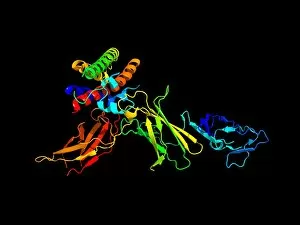
Flu virus surface protein and antibody. Computer model showing the molecular structure of the neuraminidase glycoprotein enzyme found on the surface of the influenza (flu) virus

Background imageImmune System Collection: Macrophage engulfing pathogen, artwork

Macrophage engulfing pathogen, artwork
Macrophage engulfing pathogen. Artwork of a macrophage cell (pink) engulfing a pathogen (green), a process known as phagocytosis. The pathogen will be destroyed once it has been engulfed

Background imageImmune System Collection: Immunoglobulin pentamer, artwork

Immunoglobulin pentamer, artwork
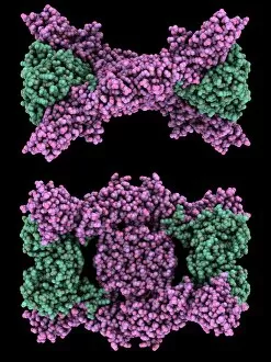
Immunoglobulin pentamer, computer artwork. Immunoglobulin (Ig) proteins, also known as antibodies, are gamma globulin proteins that are part of the mammalian immune system

Background imageImmune System Collection: Macrophage engulfing pathogens, artwork

Macrophage engulfing pathogens, artwork
Macrophage engulfing pathogens. Artwork of a macrophage cell (pink) engulfing pathogens (green), a process known as phagocytosis. The pathogens will be destroyed once they have been engulfed

Background imageImmune System Collection: Macrophage cells, TEM

Macrophage cells, TEM
Macrophage cells, coloured transmission electron micrograph (TEM). The cell nuclei are purple. Mitochondria (dark pink ovals) in the cytoplasm produce energy for the cell

Background imageImmune System Collection: Hay fever, conceptual artwork

Hay fever, conceptual artwork. Head of a human figure with flower images representing the allergic reaction known as hay fever

Background imageImmune System Collection: Lung alveoli and blood cells, TEM

Lung alveoli and blood cells, TEM
Lung alveoli and blood cells. Coloured transmission electron micrograph (TEM) of a section through lung alveoli and blood cells

Background imageImmune System Collection: Asthma and mucus overproduction, artwork

Asthma and mucus overproduction, artwork
Asthma and mucus overproduction. Artwork showing how the bodys immune response causes the inflammation seen in asthma. The chest of a human figure is at top right

Background imageImmune System Collection: Small lymphocyte, TEM

Small lymphocyte, TEM
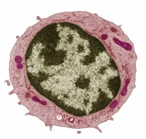
Small lymphocyte. Coloured transmission electron micrograph (TEM) showing the large central nucleus (brown) of a small lymphocyte (white blood cell)

Background imageImmune System Collection: T-cell receptor - MHC complex

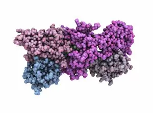
T-cell receptor - MHC complex. Computer model showing the molecular structure of a T-cell receptor (TCR, pink and blue) bound to an allogeneic (non-self) major histocompatibility complex (MHC)

Background imageImmune System Collection: White blood cell, TEM

White blood cell, TEM
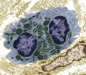
White blood cell. Coloured transmission electron micrograph (TEM) of an eosinophil (a type of white blood cell), part of the bodys immune system

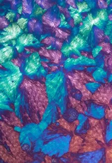
Background imageImmune System Collection: Cortisol crystals, light micrograph

Cortisol crystals, light micrograph
Cortisol crystals, polarised light micrograph. Cortisol is a steroid hormone produced by the adrenal glands, which sit on top of the kidneys

Background imageImmune System Collection: Immunoglobulin crystals, light micrograph

Immunoglobulin crystals, light micrograph
Immunoglobulin crystals, polarised light micrograph. Immunoglobulins, or antibodies, are a type of globulin protein that are produced by B cells (a type of white blood cell)

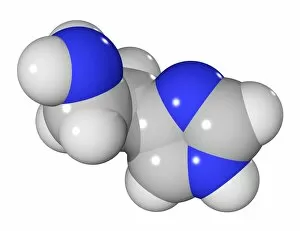
Background imageImmune System Collection: Histamine molecule

Histamine molecule. Computer model showing the structure of a molecule of histamine. Atoms are colour-coded (carbon: dark grey, hydrogen: light grey, nitrogen: blue)

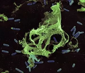
Background imageImmune System Collection: Dead neutrophil white blood cell, SEM

Dead neutrophil white blood cell, SEM
Dead neutrophil white blood cell. Coloured scanning electron micrograph (SEM) of a dead neutrophil (green) surrounded by rod-shaped bacteria (blue). Neutrophils are part of the bodys immune response

Background imageImmune System Collection: Human blood cells, SEM

Human blood cells, SEM
Human blood cells, coloured scanning electron micrograph (SEM). Seen here are platelets (small, roundish) and a neutrophil white blood cell (large, whitish)

Background imageImmune System Collection: Neutrophil engulfing TB bacteria, SEM

Neutrophil engulfing TB bacteria, SEM
Neutrophil engulfing TB bacteria. Coloured scanning electron micrograph (SEM) of a neutrophil white blood cell (large, centre) engulfing and destroying a tuberculosis (Mycobacterium tuberculosis)

Background imageImmune System Collection: Neutrophil engulfing thrush fungus, SEM

Neutrophil engulfing thrush fungus, SEM
Neutrophil engulfing thrush fungus. Coloured scanning electron micrograph (SEM) of a neutrophil white blood cell engulfing and destroying a hypha (round)

Background imageImmune System Collection: Blood cells in a kidney, SEM

Blood cells in a kidney, SEM
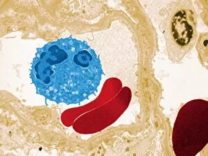
Blood cells in a kidney. Transmission electron micrograph (TEM) of a section through a kidney glomerulus, showing red blood cells (erythrocytes, red) and a white blood cell (leucocyte, blue)

Background imageImmune System Collection: Pokeroot (Phytolacca americana)

Pokeroot (Phytolacca americana)
Pokeroot flower buds (Phytolacca americana). This plant is used in herbal medicine to treat diseases related to a compromised immune system

Background imageImmune System Collection: Interleukin-12 protein molecule

Interleukin-12 protein molecule. Molecular model of a molecule of the cytokine protein interleukin-12 (IL-12). The secondary structure of the molecule is seen here

Background imageImmune System Collection: Cd28 antigen molecule

Cd28 antigen molecule. Computer model showing the secondary structure of cd28. Cd28 is an antigen found on the surface of T cells

Background imageImmune System Collection: Interleukin-10, molecular model

Interleukin-10, molecular model
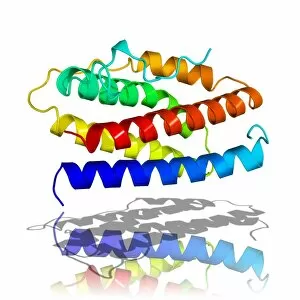
Interleukin-10. Molecular model of the secondary structure of interleukin-10, a small protein known as a cytokine that plays an important regulatory role in the bodys immune system

Background imageImmune System Collection: CD4 protein fragment, molecular model

CD4 protein fragment, molecular model
CD4 protein fragment. Molecular model of the CD4 glycoprotein, which plays a vital role in the immune system. It is found on the surface of white blood cells called T cells

Background imageImmune System Collection: Interferon, molecular model

Interferon, molecular model
Interferon. Molecular model showing the secondary structure of a molecule of interferon. Interferons are proteins produced by white blood cells as part of the immune response to invading pathogens

Background imageImmune System Collection: Interleukin-6, molecular model

Interleukin-6, molecular model
Interleukin-6. Molecular model of the secondary structure of the cytokine protein interleukin-6. This protein is produced in the body and has a wide variety of functions in the immune system

Background imageImmune System Collection: Tumour necrosis factor-alpha

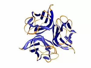
Tumour necrosis factor-alpha (TNF-alpha), computer model. This molecule is composed almost entirely of anti-parallel beta-sheets (blue)

Background imageImmune System Collection: Purple coneflowers

Purple coneflowers (Echinacea sp.) in flower. This medicinal plant can be used as a herbal treatment for wounds and burns. It is also thought to help the body to fight infections like the common cold

Background imageImmune System Collection: Purple coneflower

Purple coneflower (Echinacea sp.) flower. This medicinal plant can be used as a herbal treatment for wounds and burns. It is also thought to help the body to fight infections like the common cold

Background imageImmune System Collection: Plasma cell, TEM

Plasma cell, TEM
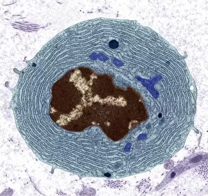
Plasma cell, coloured transmission electron micrograph (TEM). This section has revealed the cells large central nucleus (brown)


For sale as Licensed Images

Choose your image, Select your licence and Download the media

"The Immune System: A Complex Network of Defenders" Our a remarkable defense mechanism that safeguards our body against harmful invaders. At the forefront of this battle are T lymphocytes, specialized white blood cells capable of recognizing and destroying cancer cells. SEM C001 / 1679 reveals the intricate interaction between these vigilant warriors and malignant intruders. Another crucial component in our immune arsenal is the Immunoglobulin G antibody molecule, as depicted in F007 / 9894. These powerful molecules neutralize pathogens by binding to them, preventing their harmful effects on our body. Neutrophils, captured in action through SEM C018 / 8596, showcase their ability to engulf dangerous bacteria like MRSA. These fearless soldiers patrol our bloodstream, eliminating potential threats with precision and efficiency. Dendritic cells play a pivotal role in orchestrating immune responses. In an artistic representation, we see these sentinel-like cells capturing antigens and presenting them to other immune cells for recognition (artwork). The TEM image of a human white blood cell bearing HLA antigen highlights its significance in identifying foreign substances and triggering appropriate immune responses. Blood cells form the backbone of our immunity; they transport oxygen and vital nutrients while also serving as defenders against invading pathogens. Antibodies take center stage once again in captivating artwork depicting their diverse shapes and functions. They act as molecular weapons targeting specific antigens with incredible accuracy. Cortisol crystals under light micrograph remind us that stress can impact our immune system's performance. Managing stress levels becomes crucial for maintaining optimal immunity. Micrograph evidence showcases Dohle bodies within blood cells—an indicator of infection or inflammation—a reminder that vigilance is key to combating potential health threats effectively. An illustration portrays how vaccination triggers an orchestrated response involving microbes, antigens, antibodies, plasma cells (TEM), ultimately fortifying our defenses against future infections. Understanding the complexity and resilience of our immune system is essential.