
Kidney Collection (#2)

"Unveiling the Intricacies of the Kidney: A Journey through Art, Science

Background imageKidney Collection: Hunts Remedy (chromolitho)

Hunts Remedy (chromolitho)
3656998 Hunts Remedy (chromolitho) by American School, (19th century); Private Collection; (add.info.: Hunts Remedy. Illustration for trade card)

Background imageKidney Collection: Normal front view of a man showing the endocrine system including the brain

Normal front view of a man showing the endocrine system including the brain
Brain, Anatomy, Diagram, Illustration, Naked, Medical, Biology, See Through, Cut Out, White Background, Vertical, Artwork, Male Likeness, Color Image, Front View, Healthcare And Medicine

Background imageKidney Collection: PISANO, Andrea da Pontedera, called Andrea (1270-1349)

PISANO, Andrea da Pontedera, called Andrea (1270-1349). Saturn. 1337-1341. Moulding which decorated Giottos Campanile. Renaissance art. Trecento. Florentine school. Relief on rock. ITALY. Florence

Background imageKidney Collection: EyeUbiquitous_20100192

EyeUbiquitous_20100192
Food, Uncooked, Beans, Red kidney beans scattered on a white surface

Background imageKidney Collection: Advertisements at Cambridge Circus, Westminster, London, 1945

Advertisements at Cambridge Circus, Westminster, London, 1945
Advertisements at Midland Bank, Cambridge Circus, Westminster, London, 1945. Building on the south east corner covered with advertisements for Black and White whisky

Background imageKidney Collection: Bolivia Tiwanaku Site

Bolivia Tiwanaku Site
BOLIVIA. Tiahuanaco. Sculpted head, 8th-10th c. A.D. Tihuanaco culture. Sculpture on rock

Background imageKidney Collection: Cross section biomedical illustration of endocrine system in adult female

Cross section biomedical illustration of endocrine system in adult female

Background imageKidney Collection: Male anatomy of internal organs

Male anatomy of internal organs, with heart highlighted and rest of organs in half tone

Background imageKidney Collection: Male skeleton with internal organs on blue background

Male skeleton with internal organs on blue background

Background imageKidney Collection: Large staghorn calculus in the human kidney

Large staghorn calculus in the human kidney

Background imageKidney Collection: Human organs, artwork

Human organs, artwork
Human organs, computer artwork

Background imageKidney Collection: Female anatomy, historical model

Female anatomy, historical model
Female anatomy. 1920s anatomical model showing the internal organs of the female upper body with the intestines, stomach and liver removed

Background imageKidney Collection: Female abdominal anatomy, artwork

Female abdominal anatomy, artwork
Female abdominal anatomy. Computer artwork showing the kidneys and female reproductive system. Part of the lymphatic system (green) is also shown

Background imageKidney Collection: Richard Bright 1789 To 1858 British Physician Engraved By H Cook After T R Say From The Book The

Richard Bright 1789 To 1858 British Physician Engraved By H Cook After T R Say From The Book The National Portrait Gallery Volume 1 Published C1820

Background imageKidney Collection: French Advertisement C. 1916 For Urodonal A Kidney Cleanser

French Advertisement C. 1916 For Urodonal A Kidney Cleanser
French Advertisement C.1916 For Urodonal A Kidney Cleanser

Background imageKidney Collection: Entrails in the human body

Entrails in the human body.. Entrails in the human body- thyroid, stomach, esophagus, colon, pancreas, kidney, etc. Handcolored copperplate engraving from Friedrich Bertuchs Bilderbuch fur Kinder

Background imageKidney Collection: Human male anatomy

Human male anatomy
Antique illustration of a Human male anatomy

Background imageKidney Collection: Illustration of internal systems of human body

Illustration of internal systems of human body

Background imageKidney Collection: Cross section illustration of human kidney

Cross section illustration of human kidney

Background imageKidney Collection: Rear view of human skeleton showing kidney and nervous system

Rear view of human skeleton showing kidney and nervous system

Background imageKidney Collection: Nephron detail, the functional unit of excretion in the human kidney

Nephron detail, the functional unit of excretion in the human kidney

Background imageKidney Collection: Anatomy of male urinary system

Anatomy of male urinary system

Background imageKidney Collection: Cross section of internal anatomy of kidney

Cross section of internal anatomy of kidney

Background imageKidney Collection: Early stage of kidney cancer with a tumor visible on the kidney

Early stage of kidney cancer with a tumor visible on the kidney

Background imageKidney Collection: Artist depcition of abdominal aortic aneuryism (with labels)

Artist depcition of abdominal aortic aneuryism (with labels)

Background imageKidney Collection: Diagram of a nephron, the functional unit of excretion in the kidney

Diagram of a nephron, the functional unit of excretion in the kidney
Diagram of a nephron, the functional unit of excretion in the human kidney

Background imageKidney Collection: Anatomy of female body with internal organs superimposed

Anatomy of female body with internal organs superimposed

Background imageKidney Collection: Chinese acupuncture chart showing the kidney meridian

Chinese acupuncture chart showing the kidney meridian
ACUPUNCTURE CHART. Chinese acupuncture chart showing the kidney meridian

Background imageKidney Collection: Kidney stones, ultrasound scan C017 / 7381

Kidney stones, ultrasound scan C017 / 7381
Kidney stones. Ultrasound scan of a 83 year old female patient with two kidney stones (bright white) in the right kidney. Kidney stones, or calculi, are formed when mineral salts

Background imageKidney Collection: Fight-or-flight response, artwork

Fight-or-flight response, artwork
Fight-or-flight response. Artwork showing the bodys fight-or-flight response to a threat. Visual perception (yellow arrow) can trigger fear memories controlled by the brains amygdala (yellow)

Background imageKidney Collection: Male urinary system, artwork

Male urinary system, artwork
Male urinary system. Computer artwork of a male abdomen highlighting the urinary system. The two kidneys are at upper centre

Background imageKidney Collection: Polycystic kidney, ultrasound scan C017 / 7746

Polycystic kidney, ultrasound scan C017 / 7746
Polycystic kidney. Ultrasound scan of the kidney of a 42 year old female showing numerous fluid-filled cysts (dark areas)

Background imageKidney Collection: MRI scan of human abdomen showing kidneys & liver

MRI scan of human abdomen showing kidneys & liver
Abdomen. Coloured Magnetic Resonance Imaging (MRI) scan showing a coronal section through a human abdomen, front view. The lower spinal vertebrae are seen running from top to bottom through

Background imageKidney Collection: Normal abdomen, 3D CT scan

Normal abdomen, 3D CT scan
Normal abdomen. Coloured 3D computed tomography (CT) scan of the healthy abdomen and chest of a 53 year old patient. At top centre is the heart

Background imageKidney Collection: Kidney anatomy, artwork

Kidney anatomy, artwork
Kidney anatomy. Cutaway artwork showing the internal structure of a kidney. The kidney, one of a pair of such organs, is part of the human excretory system

Background imageKidney Collection: Nephron structure, artwork

Nephron structure, artwork
Nephron structure. Artwork showing the structure of a nephron, the functional filtering unit of a kidney. The function of the kidney is to filter waste products such as urea from the blood

Background imageKidney Collection: Cow anatomy, artwork

Cow anatomy, artwork
Cow anatomy. Computer artwork showing the internal anatomy of a domesticated cow (Bos primigenius). Anatomical features shown here include the skeleton, lungs, intestines, bladder

Background imageKidney Collection: Mitochondria, SEM

Mitochondria, SEM
Mitochondria. Coloured scanning electron micrograph (SEM) of mitochondria (red) in a kidney cell. Mitochondria are a type of organelle found in the cytoplasm of eukaryotic cells

Background imageKidney Collection: Illustration showing nephron structure and kidney

Illustration showing nephron structure and kidney
Human anatomy: nephron structure. Drawing

Background imageKidney Collection: Advert for Harness Electropathic corset belts 1888

Advert for Harness Electropathic corset belts 1888
Harness Electropathic belt treatment. 1888

Background imageKidney Collection: Advert for Harness Electropathic Corset Belts 1889

Advert for Harness Electropathic Corset Belts 1889
They cure nervous weakness, sleeplessness, neuralgia, debility, rheumatic affections, liver and kidney disorders, internal weakness and hysteria. 1889

Background imageKidney Collection: Advert for Harness Electropathic corset belts 1891

Advert for Harness Electropathic corset belts 1891
Advert for Harness Electropathic corset belts no taking poisonous drugs or quack nostrums. Claims to cure all rheumatic and nervous affections. 1891

Background imageKidney Collection: Seeds and their parts: garden bean 1-5, fig

Seeds and their parts: garden bean 1-5, fig 6, and kidney bean 7-9.. Handcoloured copperplate engraving of a botanical illustration by Sydenham Edwards for William Curtiss Lectures on Botany

Background imageKidney Collection: Autumnal and spring water starworts, kidney-leaved sowerweed

Autumnal and spring water starworts, kidney-leaved sowerweed.. Handcolored botanical copperplate engraving from Joshua Hamiltons Culpepers English Family Physician; or Medical Herbal Enlarged

Background imageKidney Collection: Milestone (2nd century BC). It comes from Santa

Milestone (2nd century BC). It comes from Santa Eulalia de Ron确 a (Barcelona). Roman art. Sculpture. SPAIN. CATALONIA. BARCELONA. Barcelona. Archaeology Museum of Catalonia

Background imageKidney Collection: Head. Iron Age

Head. Iron Age
Head from Khirbet el Hajjar (10th c. BC). Iron Age. Sculpture on rock. JORDAN. Amman. Archaeological Museum

Background imageKidney Collection: Head of man. Iron Age

Head of man. Iron Age
Head of man. 9th-5th c. BC. Sculpture with Egyptian influence. Iron Age. Sculpture on rock. JORDAN. Amman. Archaeological Museum

Background imageKidney Collection: Picture No. 11806843

Picture No. 11806843
hematite, botryoidal form, kidney ore, Cumberland, England hematite. Date:


For sale as Licensed Images

Choose your image, Select your licence and Download the media

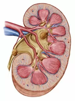
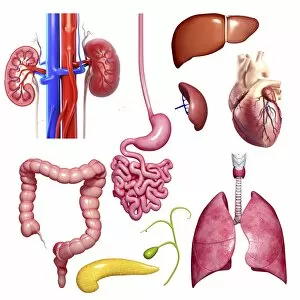
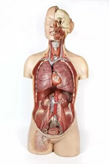
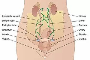
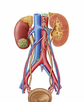
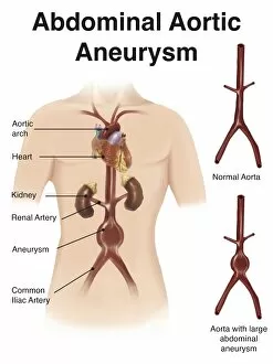
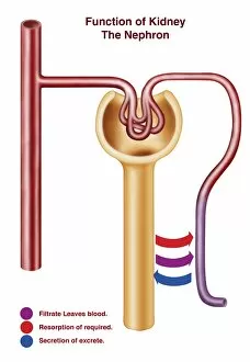
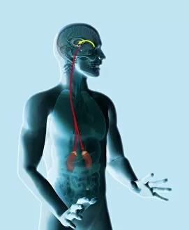
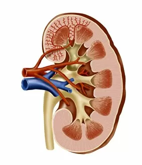
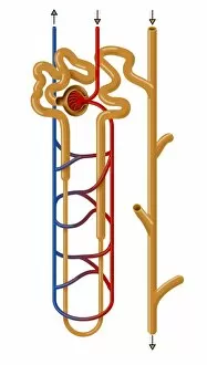
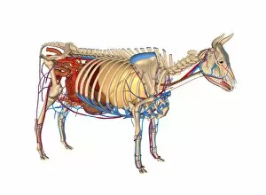
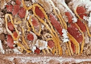
"Unveiling the Intricacies of the Kidney: A Journey through Art, Science, and History" Embarking on a full body scan or an MRI scan reveals the hidden wonders within our bodies. As we delve deeper into these scans, we discover the intricate network of our cardiovascular system and witness its harmonious dance with other organs. Throughout history, artists have captured the essence of life in their artwork. From ancient masterpieces to modern illustrations, they have depicted various aspects of human anatomy. One such illustration showcases internal organs like the heart, lung, intestines, pancreas, kidney, and even testis in a snake—a testament to nature's complexity. Zooming closer into microscopic details brings us face-to-face with kidney tubules in section—an awe-inspiring sight that highlights their vital role in maintaining our well-being. Just as Tsar Alexander III cherished his family's health and happiness during his reign; so too do our kidneys work tirelessly to ensure our bodies function optimally. In a color MRI scan of the abdomen showcasing kidneys and liver side by side—two unsung heroes—we are reminded of their significance for overall health. Spare kidneys stand as silent guardians ready to step up if needed—nature's incredible backup plan ensuring resilience against adversity. Stepping back into ancient times again takes us to Egyptian art where statues like Djoser reveal reverence for bodily functions including those performed by kidneys—a symbol of vitality and strength. Meanwhile, red kidney beans remind us that nature provides nourishment not only for sustenance but also for organ support. Delving further into scientific exploration leads us to marvel at intricate structures like kidney glomerulus when viewed under scanning electron microscopy (SEM). These mesmerizing images showcase nature's attention to detail while reminding us how every tiny element contributes towards optimal functioning. Lastly, X-ray images capturing abdominal arteries give insight into blood flow dynamics—the lifeline connecting all parts of our body including vital organs like the kidneys.