Kidney Collection (#4)

"Unveiling the Intricacies of the Kidney: A Journey through Art, Science

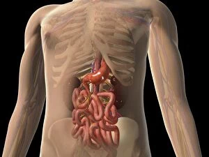
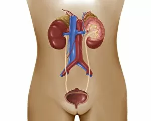
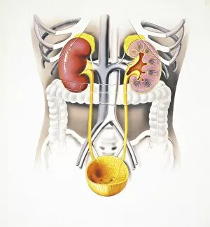
Background imageKidney Collection: Transparent view of human body showing kidney and intestines

Transparent view of human body showing kidney and intestines

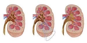
Background imageKidney Collection: Comparison of different sized kidney stones in the human kidney

Comparison of different sized kidney stones in the human kidney. The middle image shows the use of extracorporeal shock wave lithotripsy to break up the stones

Background imageKidney Collection: Hydronephrosis condition of the kidney

Hydronephrosis condition of the kidney
Hydronephrosis is a condition that typically occurs when one kidney becomes swollen due to the failure of normal drainage of urine from the kidney to the bladder

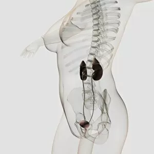
Background imageKidney Collection: Three dimensional view of female urinary system

Three dimensional view of female urinary system, profile view

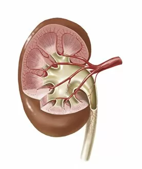
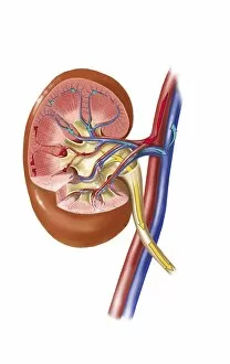
Background imageKidney Collection: Human kidney with artery

Human kidney with artery

Background imageKidney Collection: Cutaway view of the human kidney and adrenal gland

Cutaway view of the human kidney and adrenal gland

Background imageKidney Collection: Anatomy of female urinary system

Anatomy of female urinary system

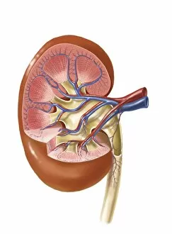
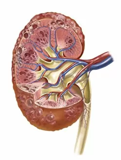
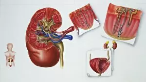
Background imageKidney Collection: Interior detail of human kidney

Interior detail of human kidney

Background imageKidney Collection: Human skeleton showing kidney, stomach and intestines

Human skeleton showing kidney, stomach and intestines
Human skeleton showing kidney, stomach, intestines and nervous system

Background imageKidney Collection: Medical chart showing the signs and symptoms of kidney stones

Medical chart showing the signs and symptoms of kidney stones

Background imageKidney Collection: Renal Corpuscle and the filtration membrane

Renal Corpuscle and the filtration membrane. Glomerular capillary surrounded by podocytes

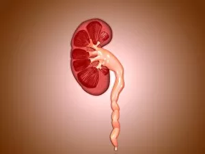
Background imageKidney Collection: Conceptual image of kidneys with ureter

Conceptual image of kidneys with ureter

Background imageKidney Collection: Human skeleton showing kidneys and nervous system

Human skeleton showing kidneys and nervous system

Background imageKidney Collection: Human urinary system

Human urinary system

Background imageKidney Collection: Urine production in human kidney

Urine production in human kidney

Background imageKidney Collection: Human kidney

Human kidney

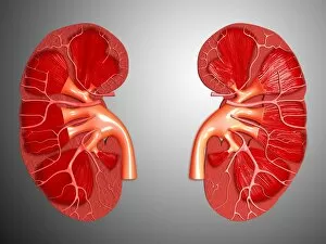
Background imageKidney Collection: Cross-section of human kidney

Cross-section of human kidney

Background imageKidney Collection: Polycystic kidney

Polycystic kidney

Background imageKidney Collection: Ureterovesical junction (UVJ) in the kidney

Ureterovesical junction (UVJ) in the kidney. This is the point where the ureters from the kidney connect to the bladder

Background imageKidney Collection: Anatomy of adrenal gland, transverse section

Anatomy of adrenal gland, transverse section

Background imageKidney Collection: Human kidney and adrenal gland

Human kidney and adrenal gland

Background imageKidney Collection: Peak Kidney, Jan Gillisz. van Vliet, 1635

Peak Kidney, Jan Gillisz. van Vliet, 1635

Background imageKidney Collection: Pikeman, Raphael de Mey, Hendrick Goltzius, Johann Bussemacher, 1580 - 1600

Pikeman, Raphael de Mey, Hendrick Goltzius, Johann Bussemacher, 1580 - 1600

Background imageKidney Collection: Picture No. 11073985

Picture No. 11073985
Flowery Meadow with Kidney Vetch and Winged Broom Piatra Craiului National Park, Southern Carpathians, Romania

Background imageKidney Collection: Picture No. 10947868

Picture No. 10947868
Porth Kidney - view looking to Godrevy Cornwall, UK. Date:

Background imageKidney Collection: Picture No. 10886848

Picture No. 10886848
Kidney-vetch - in flower in masses (Anthyllis vulneraria) Date:

Background imageKidney Collection: Picture No. 10886821

Picture No. 10886821
Thrift - and Kidney Vetch in May (Armeria maritima) Date:

Background imageKidney Collection: Picture No. 10886811

Picture No. 10886811
Kidney Vetch - masses in spring (Anthyllis vulneraria) Date:

Background imageKidney Collection: Picture No. 10876992

Picture No. 10876992
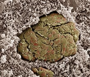
Scanning electron micrograph (SEM) of Kidney glomerulus Date:

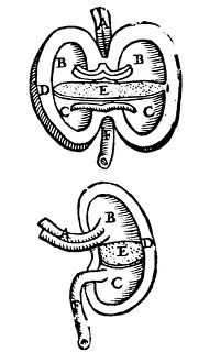
Background imageKidney Collection: Woodcut disproving the then current notion that the pelvis of the kidney was divided by a sieve or

Woodcut disproving the then current notion that the pelvis of the kidney was divided by a sieve or filter E
VESALIUS: KIDNEYS, 1543. Woodcut disproving the then current notion that the pelvis of the kidney was divided by a sieve or filter E, into two chambers

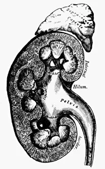
Background imageKidney Collection: Cross-section of a kidney. Wood engraving, English, late 19th century

Cross-section of a kidney. Wood engraving, English, late 19th century
HUMAN KIDNEY. Cross-section of a kidney. Wood engraving, English, late 19th century

Background imageKidney Collection: Hydrotherapy treatments. Wet packs for treating the the kidney (top) and spine (middle)

Hydrotherapy treatments. Wet packs for treating the the kidney (top) and spine (middle). Bottom: sitz-bath and half-bath. One of the oldest forms of medical treatment

Background imageKidney Collection: Illustration showing kidney

Illustration showing kidney
Medicine: kidney

Background imageKidney Collection: Illustration showing excretory organs

Illustration showing excretory organs
Medicine: excretory organs

Background imageKidney Collection: Illustration of abdomen with intestinal organs and kidneys

Illustration of abdomen with intestinal organs and kidneys
Medicine: abdomen with intestinal organs and kidneys. Drawing
Background imageKidney Collection: Medical illustration of Urinary System with kidney, drawing

Medical illustration of Urinary System with kidney, drawing
Medicine - Anatomy - Urinary System - Kidney. Drawing

Background imageKidney Collection: Illustration of kidney specimen

Illustration of kidney specimen
Medicine: Human anatomy, kidney specimen

Background imageKidney Collection: Illustration of position of the kidney in human body

Illustration of position of the kidney in human body
Medicine: Human anatomy, position of the kidney in the body. Drawing

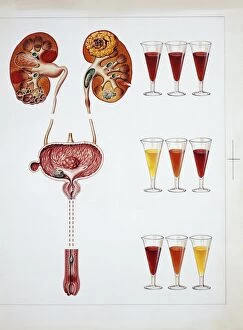
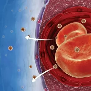
Background imageKidney Collection: Hematuria: presence of red blood cells in urinary system, drawing

Hematuria: presence of red blood cells in urinary system, drawing
Medicine - Anatomy - Urinary System - Hematuria: presence of red blood cells in the urine. Drawing

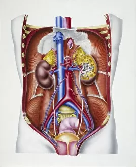
Background imageKidney Collection: Urinary System and Endocrine system, kidneys and adrenal glands with their vascular supply, drawing

Urinary System and Endocrine system, kidneys and adrenal glands with their vascular supply, drawing
Medicine - Anatomy - Urinary System - Kidney - Endocrine system - Adrenal glands - Kidneys and adrenal glands with their vascular supply. Drawing

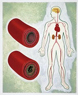
Background imageKidney Collection: Atherosclerosis, drawing

Atherosclerosis, drawing
Medicine - Pathology - Atherosclerosis, drawing

Background imageKidney Collection: Digital composite showing process of removing waste from human kidney using haemodialysis

Digital composite showing process of removing waste from human kidney using haemodialysis

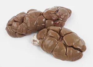
Background imageKidney Collection: Veal kidneys against white background

Veal kidneys against white background

Background imageKidney Collection: Using fingers to peel membrane off calfs kidney

Using fingers to peel membrane off calfs kidney

Background imageKidney Collection: Pulling away white fat (suet) surrounding whole kidney

Pulling away white fat (suet) surrounding whole kidney

Background imageKidney Collection: Peeling off the outer membrane of lambs kidney, close-up

Peeling off the outer membrane of lambs kidney, close-up

Background imageKidney Collection: Veal kidney being fried in oil in a heavy frying pan

Veal kidney being fried in oil in a heavy frying pan


For sale as Licensed Images

Choose your image, Select your licence and Download the media

"Unveiling the Intricacies of the Kidney: A Journey through Art, Science, and History" Embarking on a full body scan or an MRI scan reveals the hidden wonders within our bodies. As we delve deeper into these scans, we discover the intricate network of our cardiovascular system and witness its harmonious dance with other organs. Throughout history, artists have captured the essence of life in their artwork. From ancient masterpieces to modern illustrations, they have depicted various aspects of human anatomy. One such illustration showcases internal organs like the heart, lung, intestines, pancreas, kidney, and even testis in a snake—a testament to nature's complexity. Zooming closer into microscopic details brings us face-to-face with kidney tubules in section—an awe-inspiring sight that highlights their vital role in maintaining our well-being. Just as Tsar Alexander III cherished his family's health and happiness during his reign; so too do our kidneys work tirelessly to ensure our bodies function optimally. In a color MRI scan of the abdomen showcasing kidneys and liver side by side—two unsung heroes—we are reminded of their significance for overall health. Spare kidneys stand as silent guardians ready to step up if needed—nature's incredible backup plan ensuring resilience against adversity. Stepping back into ancient times again takes us to Egyptian art where statues like Djoser reveal reverence for bodily functions including those performed by kidneys—a symbol of vitality and strength. Meanwhile, red kidney beans remind us that nature provides nourishment not only for sustenance but also for organ support. Delving further into scientific exploration leads us to marvel at intricate structures like kidney glomerulus when viewed under scanning electron microscopy (SEM). These mesmerizing images showcase nature's attention to detail while reminding us how every tiny element contributes towards optimal functioning. Lastly, X-ray images capturing abdominal arteries give insight into blood flow dynamics—the lifeline connecting all parts of our body including vital organs like the kidneys.