
Knee Cap Collection
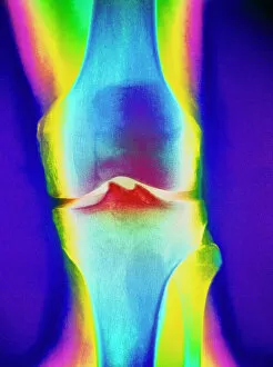
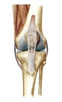
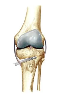
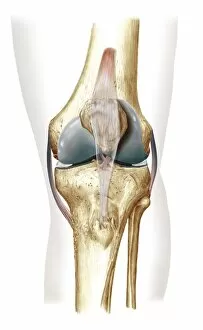
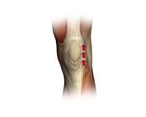
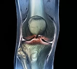
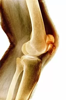
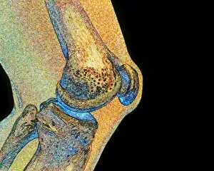
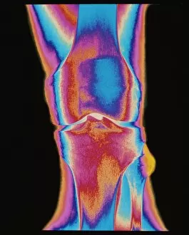
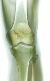
"Unveiling the Intricacies of the Knee Cap: A Fascinating Journey into Joint Health" In this captivating image, a coloured X-ray of a human knee joint takes center stage


For sale as Licensed Images
Choose your image, Select your licence and Download the media
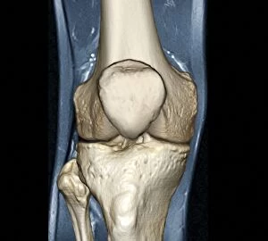
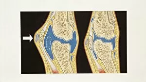
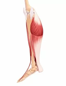
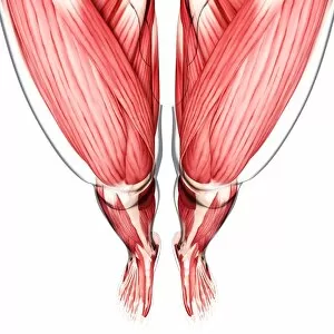
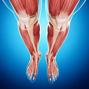
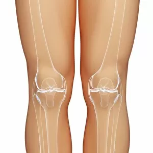
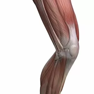
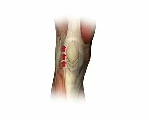
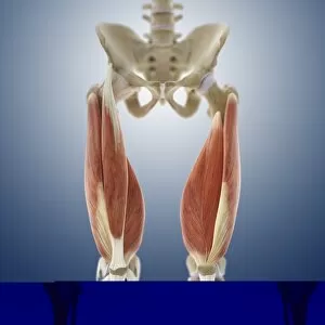
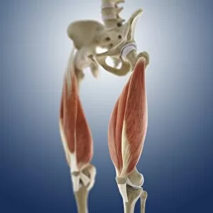
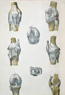
"Unveiling the Intricacies of the Knee Cap: A Fascinating Journey into Joint Health" In this captivating image, a coloured X-ray of a human knee joint takes center stage. The intricate details reveal a healthy knee, as depicted in the CT scan C018 / 0413. Accompanying this visual is an illustration showcasing the biceps brachii muscle, emphasizing the interconnectedness between different parts of our body. As we explore further, we encounter illustrations highlighting human bones such as the patella or knee cap through ballottement. The artwork depicting human leg musculature in various forms - F007 / 1930, F007 / 1929, and F007 / 1599 - adds depth to our understanding of how these muscles contribute to overall leg strength and mobility. Delving deeper into knee anatomy through additional artwork reveals even more intricacies within this remarkable joint. Artwork like F007 / 1212 showcases specific aspects while other pieces like F007 / 1003 and F007 / 0978 provide comprehensive views that leave us awe-inspired by its complexity. This collection serves as a reminder that our knees are not just simple hinges but rather sophisticated structures composed of bones, muscles, tendons, and ligaments working harmoniously together to support movement and bear weight. Let us marvel at these incredible images that shed light on the wonders of our knee cap – an essential component enabling us to walk tall and embrace life's adventures with confidence.