Home > Popular Themes > Human Body
Knee muscles and bones, artwork C016 / 7013

![]()
Wall Art and Photo Gifts from Science Photo Library
Knee muscles and bones, artwork C016 / 7013
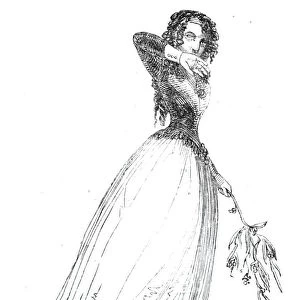
Knee muscles and bones. Artwork of an oblique lateral frontal view of the muscles and bones of a human knee. This view is of the outside of the left leg, with the iliotibial band (white) running down the outside of the thigh and attaching to Gerdys tubercle on the outside of the tibia (one of the lower leg bones). The femur (thigh bone) and patella (knee-cap) are visible below the muscles. Muscles, ligaments and tendons include the patellar ligament and the quadriceps femoris, vastus lateralis, and vastus medialis obliquus thigh muscles (upper right). In the lower leg, the calf muscles are the gastrocnemius and the soleus (lower centre)
Science Photo Library features Science and Medical images including photos and illustrations
Media ID 9245891
© D & L GRAPHICS / SCIENCE PHOTO LIBRARY
Anterior Bones Femur Frontal Gastrocnemius Iliotibial Band Joint Knee Knee Cap Kneecap Lateral Ligament Ligaments Lower Leg Muscles Oblique Patella Patellar Ligament Quadriceps Femoris Soleus Tendon Tendons Thigh Tibia Vastus Lateralis Iliotibial Tract
EDITORS COMMENTS
This artwork, titled "Knee muscles and bones" offers a mesmerizing glimpse into the intricate anatomy of the human knee. With an oblique lateral frontal view, it showcases the muscles and bones that make up this vital joint. The focus is on the outside of the left leg, where we can observe the iliotibial band gracefully running down the thigh and attaching to Gerdys tubercle on the tibia. The femur, our sturdy thigh bone, and patella or knee-cap are also visible below these muscular structures. Amongst them, we can identify several crucial components such as ligaments and tendons like the patellar ligament. In addition to this, prominent thigh muscles like quadriceps femoris, vastus lateralis, and vastus medialis obliquus take their place in this artistic representation. Moving towards lower leg anatomy within this illustration reveals two significant calf muscles - gastrocnemius and soleus - positioned at its center. Against a white background that accentuates every detail with clarity, this artwork provides a comprehensive visual understanding of knee structure. With its emphasis on normal anatomical features associated with a healthy joint function, this print serves as an invaluable resource for biology enthusiasts or medical professionals seeking to deepen their knowledge about knees' complexity. Created by D & L GRAPHICS for Science Photo Library's collection of scientific illustrations; it stands as both visually stunning artistry and educational tool in one frame.
MADE IN THE USA
Safe Shipping with 30 Day Money Back Guarantee
FREE PERSONALISATION*
We are proud to offer a range of customisation features including Personalised Captions, Color Filters and Picture Zoom Tools
SECURE PAYMENTS
We happily accept a wide range of payment options so you can pay for the things you need in the way that is most convenient for you
* Options may vary by product and licensing agreement. Zoomed Pictures can be adjusted in the Cart.