
Laboratory Equipment Collection (#6)

"Exploring the World of Science

Background imageLaboratory Equipment Collection: Legionella bacteria culture

Legionella bacteria culture
MODEL RELEASED. Legionella bacteria culture. Microbiologist holding a petri dish containing a culture of Legionella sp. bacteria. Legionella sp

Background imageLaboratory Equipment Collection: Cultured Staphylococcus bacteria

Cultured Staphylococcus bacteria
Staphylococcus bacteria. Cultured colonies of Staphylococcus aureus bacteria in a petri dish. S. aureus is a Gram-positive, non- motile

Background imageLaboratory Equipment Collection: Bacillus cereus food poisoning

Bacillus cereus food poisoning
MODEL RELEASED. Bacillus cereus food poisoning. Microbiologist holding a petri dish containing a culture of B. cereus bacteria in one hand and a sample of reheated rice in the other. B

Background imageLaboratory Equipment Collection: Listeria culture

Listeria culture
MODEL RELEASED. Listeria culture. Microbiologist holding a petri dish containing a culture of Listeria monocytogenes bacteria

Background imageLaboratory Equipment Collection: Bacillus cereus culture

Bacillus cereus culture
MODEL RELEASED. Bacillus cereus culture. Microbiologist holding a petri dish containing a culture of B. cereus bacteria. B

Background imageLaboratory Equipment Collection: Bacterial colonies

Bacterial colonies. Petri dish containing colonies of bacteria cultured from a hand print found in a house. The print was lifted from a surface on a growth medium gel

Background imageLaboratory Equipment Collection: View of a petri dish with bacterial cultures

View of a petri dish with bacterial cultures

Background imageLaboratory Equipment Collection: Bacteria water test

Bacteria water testing. Female researcher removing a membrane filter from a bacterial culture dish with forceps as she tests a water sample for bacterial contamination

Background imageLaboratory Equipment Collection: Gloved hand of a researcher holding a petri dish

Gloved hand of a researcher holding a petri dish
Petri dish. Gloved hand of a researcher using forceps to place a grid on an agar plate in order to grow colonies of bacteria as part of a water purity check

Background imageLaboratory Equipment Collection: Technician with bacterial cultures in petri dishes

Technician with bacterial cultures in petri dishes
MODEL RELEASED. Bacterial cultures. Masked techni- cian holding two petri dishes containing bacterial cultures in gloved hands

Background imageLaboratory Equipment Collection: Stack of petri dishes with bacterial cultures

Stack of petri dishes with bacterial cultures
Bacterial cultures. View of a stack of petri dishes containing bacterial cultures. These petri dishes are filled with different coloured agar nutrient media on which bacteria are grown

Background imageLaboratory Equipment Collection: Petri dishes containing bacterial cultures

Petri dishes containing bacterial cultures
Bacterial culture. View of petri dishes containing bacterial cultures. These petri dishes are filled with different coloured agar nutrient media on which bacteria are grown

Background imageLaboratory Equipment Collection: Close-up of petri dishes with bacterial cultures

Close-up of petri dishes with bacterial cultures
Bacterial culture. Close-up of petri dishes containing bacterial cultures. These petri dishes are filled with different coloured agar nutrient media on which bacteria are grown

Background imageLaboratory Equipment Collection: Hands with petri dishes with Aspergillus cultures

Hands with petri dishes with Aspergillus cultures

Background imageLaboratory Equipment Collection: Researcher testing the sensitivity of bacteria

Researcher testing the sensitivity of bacteria
Model released. Researcher testing the sensitivity of bacteria on culture plates. The plunger-like device which he is using is able to dispense six small portions of different antibiotics

Background imageLaboratory Equipment Collection: Quality control on media to grow microorganisms

Quality control on media to grow microorganisms
Model released. Researchers testing the performance of different media used to grow microorganisms such as bacteria. The image was taken in one of the 52 laboratories found throughout the UK forming

Background imageLaboratory Equipment Collection: Researcher & samples

Researcher & samples
Model released. Researcher removing trays containing samples of bacteria which were stored at low temperature in liquid nitrogen

Background imageLaboratory Equipment Collection: Researchers handling trays of frozen bacteria

Researchers handling trays of frozen bacteria
Model released. Researchers removing trays containing samples of bacteria which were stored at low temperature in liquid nitrogen

Background imageLaboratory Equipment Collection: Different media used to store or grow bacteria

Different media used to store or grow bacteria
Bacteria in culture. Different media used to grow or store cultures of bacteria. At centre some Petri dishes are seen in which bacteria grow in agar

Background imageLaboratory Equipment Collection: Searching for bacteria in frozen food

Searching for bacteria in frozen food

Background imageLaboratory Equipment Collection: Media use for growing or storing bacteria

Media use for growing or storing bacteria
Bacteria in culture. Different media used to grow or store cultures of bacteria. At centre some Petri dishes are seen in which bacteria grow in agar

Background imageLaboratory Equipment Collection: Agar filling machine

Agar filling machine
MODEL RELEASED. Agar filling machine. Technician lifts filled petri dishes from an agar machine. Empty petri dishes are at upper left. Agar is a nutrient medium upon which bacteria can be grown

Background imageLaboratory Equipment Collection: Filter paper, SEM

Filter paper, SEM
Filter paper. Scanning electron micrograph (SEM) of the cut end of a piece of filter paper showing its many cellulose fibres

Background imageLaboratory Equipment Collection: Used Eppendorf tubes

Used Eppendorf tubes

Background imageLaboratory Equipment Collection: Multiwell sample tray

Multiwell sample tray

Background imageLaboratory Equipment Collection: Pipetting

Pipetting
MODEL RELEASED. Pipetting liquid into an Eppendorf tube

Background imageLaboratory Equipment Collection: Petri dish bacterial culture being placed in stack

Petri dish bacterial culture being placed in stack
MODEL RELEASED. Bacterial cultures. Gloved hand places a petri dish with a bacterial culture back onto a petri dish stack

Background imageLaboratory Equipment Collection: Electrical circuit

Electrical circuit. This circuit consists of three light bulbs lit by a 6-volt power supply. The power supply comprises four 1.5-volt batteries connected in series

Background imageLaboratory Equipment Collection: Orange battery

Orange battery. Copper (orange) and zinc (silver) electrodes have been inserted into an orange and connected to wires to produce electricity. The voltmeter (top) is showing the voltage generated

Background imageLaboratory Equipment Collection: Tomato battery

Tomato battery
Lemon battery. Copper (orange) and zinc (silver) electrodes have been inserted into a lemon and connected to wires to produce electricity. The voltmeter (top) is showing the voltage generated

Background imageLaboratory Equipment Collection: Digital pH measurement

Digital pH measurement

Background imageLaboratory Equipment Collection: Medical blood analysis

Medical blood analysis. Glass slides being used in a hospital laboratory to analyse a blood sample from a patient, to help diagnose diseases. Photographed at St. Marys Hospital in Lacor, Gulu, Uganda

Background imageLaboratory Equipment Collection: Stem cell research

Stem cell research
MODEL RELEASED. Stem cell research. Scientist examining stem cell cultures

Background imageLaboratory Equipment Collection: Biological research

Biological research
MODEL RELEASED. Biological research. Scientist using a pipette to fill eppendorf vials. These are used in a wide range of biological experiments, ranging from genetic analysis to medical research

Background imageLaboratory Equipment Collection: Microbiological research

Microbiological research
MODEL RELEASED. Microbiological research. Scientist examining microbiological cultures in a petri dish

Background imageLaboratory Equipment Collection: Bacterial culture

Bacterial culture. Petri dish containing bacterial cultures (red). The bacteria are grown on agar jelly (beige), which provides all the nutrients needed by bacteria for growth


For sale as Licensed Images

Choose your image, Select your licence and Download the media

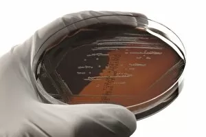
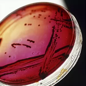
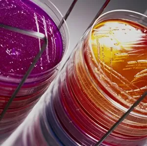
"Exploring the World of Science: A Journey through Laboratory Equipment" Step into the world of scientific discovery as we delve into the fascinating realm of laboratory equipment. From groundbreaking experiments to revolutionary discoveries, these tools have played a pivotal role in shaping our understanding of the natural world. In 1935, French scientists Frederic Joliot and Irene Joliot-Curie paved the way for nuclear chemistry with their pioneering research. Their work laid the foundation for future advancements in this field, forever changing our perception of atomic structure. The importance of maintaining a safe environment cannot be overstated, especially when dealing with hazardous substances like Salmonella culture or bacterial culture. Laboratory equipment ensures that researchers can handle these materials safely while studying their properties and effects. Microscopes have been instrumental in unraveling some of nature's most intricate secrets. Focus stacked images reveal an inverted wonderland on microscope slides - diatoms. These single-celled algae not only produce a significant portion of Earth's oxygen but also play a crucial role in global carbon fixation. Throughout history, notable figures have made indelible contributions to science. Antoine Laurent Lavoisier and his wife were immortalized by Jacques-Louis David in 1788, symbolizing their dedication to chemical analysis and establishing modern chemistry as we know it today. Artistic depictions often capture moments frozen in time, shedding light on scientific practices from centuries past. The frontispiece for the Society of Etchers from 1863 showcases Paris' Municipal Chemical Laboratory - a hub where innovative minds converged to push boundaries and unlock new knowledge. David Wiley's portrait from around 1801 portrays him engrossed in his scientific pursuits - a testament to the unwavering curiosity that drives researchers forward. Similarly, "Man of Science" (1839) encapsulates the spirit of exploration shared by countless individuals dedicated to expanding human knowledge. "In the Laboratory" (ca.