Leucocytes Collection (#3)

"Exploring the Intricate World of Leucocytes: A Journey through Blood Coagulation Cascade and Cellular Artwork" In this captivating caption

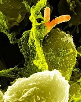
Background imageLeucocytes Collection: Neutrophils and Shigella bacteria, SEM

Neutrophils and Shigella bacteria, SEM
Neutrophils and Shigella bacteria. Coloured scanning electron micrograph (SEM) of neutrophil white blood cells (large, yellow-green) and Shigella bacteria (orange, upper right)

Background imageLeucocytes Collection: Blood cells, computer artwork

Blood cells, computer artwork
Blood cells. Computer artwork of red blood cells, white blood, white blood cells (blue) and platelets (yellow) travelling through the lumen of the blood vessel

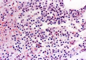
Background imageLeucocytes Collection: Human blood cells, light micrograph

Human blood cells, light micrograph
Human blood cells. Light micrograph of blood cells at a site of inflammation, showing red blood cells (erythrocytes, red) surrounded by many granulocytes (blue)

Background imageLeucocytes Collection: Coronary thrombosis, light micrograph

Coronary thrombosis, light micrograph
Coronary thrombosis. Light micrograph of a section through a thrombus (blood clot) in the coronary artery of the heart. The clot is packed full of red blood cells (red)

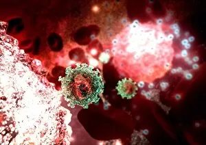
Background imageLeucocytes Collection: Budding HIV particle, computer artwork

Budding HIV particle, computer artwork
Budding HIV particle. Computer artwork of an HIV (human immunodeficiency virus) particle (green) exiting a T-lymphocyte (a type of white blood cell, lower left)

Background imageLeucocytes Collection: Artery, TEM

Artery, TEM
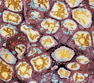
Artery. Coloured transmission electron micrograph (TEM) of a transverse section through an artery. Red blood cells (erythrocytes, red) and white blood cells (leucocytes)

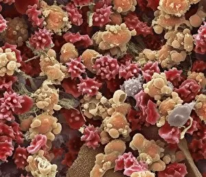
Background imageLeucocytes Collection: Cells from a urine infection, SEM

Cells from a urine infection, SEM
Cells from a urine infection. Coloured scanning electron micrograph (SEM) of crenated red blood cells, white blood cells and epithelial cells present in a urine sample taken from someone with a

Background imageLeucocytes Collection: Activated granulocytes, SEM

Activated granulocytes, SEM
Activated granulocytes. Coloured scanning electron micrograph (SEM) of activated granulocyte, or polymorphonucleocytes (PMNs), white blood cells

Background imageLeucocytes Collection: Monocyte white blood cells, artwork

Monocyte white blood cells, artwork
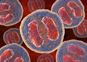
Monocyte white blood cells. Computer artwork of monocyte white blood cells, showing their lobed nuclei (purple, centre). Monocytes, like all white blood cells

Background imageLeucocytes Collection: Basophil white blood cells, artwork

Basophil white blood cells, artwork
Basophil white blood cells. Computer artwork of basophil white blood cells, showing their lobed nuclei (red). Basophils are the smallest and least common of the white blood cells

Background imageLeucocytes Collection: Lymph node, TEM

Lymph node, TEM
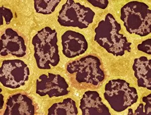
Lymph node. Coloured transmission electron micrograph (TEM) of a section through the peripheral corona of a lymph nodule, showing numerous small lymphocyte white blood cells (dark)

Background imageLeucocytes Collection: Eosinophil white blood cells, artwork

Eosinophil white blood cells, artwork
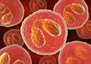
Eosinophil white blood cells. Computer artwork of eosinophil white blood cells, showing their lobed nuclei (centre). Eosinophils, like all white blood cells, are part of the immune system

Background imageLeucocytes Collection: Immune system, artwork

Immune system, artwork
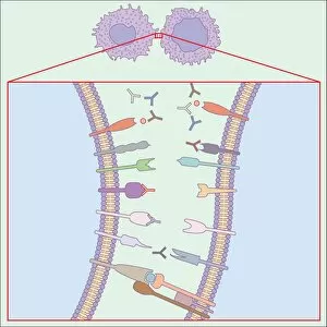
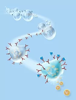
Immune system. Computer artwork showing how T-cells (right), antibodies (Y-shaped) and antigen-presenting cells (APC, left) interact during an immune response

Background imageLeucocytes Collection: Human immune response, artwork

Human immune response, artwork
Human immune response. Computer artwork showing the interactions of the various white blood cells (purple) involved during an immune response in the human body

Background imageLeucocytes Collection: Immune response to chronic inflammation

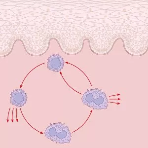
Immune response to chronic inflammation. Computer artwork showing the interaction of T cell, lymphocyte, and macrophage white blood cells in response to chronic inflammation of the skin (top)

Background imageLeucocytes Collection: Skin damage immune response, artwork

Skin damage immune response, artwork
Skin damage immune response. Computer artwork showing the bodys immune response to pathogens (red dots) entering through a skin wound (upper centre)

Background imageLeucocytes Collection: Granulocytes, SEM

Granulocytes, SEM
Granulocytes. Coloured scanning electron micrograph (SEM) of granulocytes in bone marrow. Granulocytes are a category of white blood cell characterized by the presence of granules in their cytoplasm

Background imageLeucocytes Collection: Brain cells, light micrograph

Brain cells, light micrograph
Brain cells. Fluorescence light micrograph of activated microglial cells (stained for membrane co-factor protein, yellow) and oligodendrocytes (stained for myelin basic protein, red)

Background imageLeucocytes Collection: Allergic response, artwork

Allergic response, artwork
Allergic response. Computer artwork showing the process that produces an allergic reaction. Antigens (blue), such as pollen

Background imageLeucocytes Collection: Spleen cell, TEM

Spleen cell, TEM
Spleen cell. Coloured transmission electron micrograph (TEM) of a section through a cell in the spleen, an organ located in the upper left side of the abdomen in vertebrate animals

Background imageLeucocytes Collection: Spleen cells, TEM

Spleen cells, TEM
Spleen cells. Coloured transmission electron micrograph (TEM) of a section through cells in the spleen, an organ located in the upper left side of the abdomen in vertebrate animals

Background imageLeucocytes Collection: Lung alveoli and blood cells, TEM

Lung alveoli and blood cells, TEM
Lung alveoli and blood cells. Coloured transmission electron micrograph (TEM) of a section through lung alveoli and blood cells

Background imageLeucocytes Collection: Blood cells in blood vessel, artwork

Blood cells in blood vessel, artwork
Blood cells in blood vessel, computer artwork. Red blood cells (erythrocytes, red) distribute oxygen to body tissues, and carry waste carbon dioxide back to the lungs

Background imageLeucocytes Collection: White blood cell, TEM

White blood cell, TEM
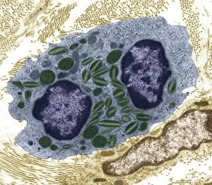
White blood cell. Coloured transmission electron micrograph (TEM) of an eosinophil (a type of white blood cell), part of the bodys immune system

Background imageLeucocytes Collection: Human blood cells, SEM

Human blood cells, SEM
Human blood cells, coloured scanning electron micrograph (SEM). Seen here are platelets (small, roundish) and a neutrophil white blood cell (large, whitish)

Background imageLeucocytes Collection: Going to the Attack

Going to the Attack
Going to the attack A depiction of leucocytes and phagocytes as the Army of the Interior, armed with bows and arrows, on their way to fight off infection


For sale as Licensed Images

Choose your image, Select your licence and Download the media

"Exploring the Intricate World of Leucocytes: A Journey through Blood Coagulation Cascade and Cellular Artwork" In this captivating caption, we delve into the fascinating realm of leucocytes, also known as white blood cells. These microscopic warriors play a crucial role in our immune system's defense against harmful invaders. The journey begins with artwork C016/9873, which depicts the intricate process of the blood coagulation cascade. This mesmerizing image showcases how leucocytes work alongside other components to form clots and prevent excessive bleeding. Moving on to scanning electron microscopy (SEM), we witness the beauty of red and white blood cells in stunning detail. SEM images such as C016/3099 and C016/3098 showcase these cellular marvels side by side with platelets, highlighting their collective effort in maintaining our health. Transitioning to transmission electron microscopy (TEM), plasma cells take center stage in TEM imagery. These specialized white blood cells are responsible for producing antibodies that target specific pathogens, safeguarding us from infections. The TEM image reveals their unique structure and function (TEM). Next up is an activated macrophage captured through SEM (C015/6375). This powerful defender engulfs foreign particles or damaged cells, playing a critical role in immune responses. Artwork showcasing lymphocyte white blood cells reminds us of their diversity and importance within our immune system's arsenal. Each type has its own distinct function but works together harmoniously to protect our bodies from harm. As we explore further under SEM lenses, monocyte white blood cell (SEM C016 / 3089) comes into view – another versatile player capable of transforming into different types of immune defenders when needed. No exploration would be complete without acknowledging human red blood cells' essential role in transporting oxygen throughout our bodies efficiently - beautifully showcased via SEM imagery. Finally, multiple SEM images highlight various aspects of general "blood cell" morphology, emphasizing the intricate beauty within our circulatory system.