
Leucocytes Collection (#2)
"Exploring the Intricate World of Leucocytes: A Journey through Blood Coagulation Cascade and Cellular Artwork" In this captivating caption


For sale as Licensed Images
Choose your image, Select your licence and Download the media
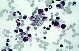
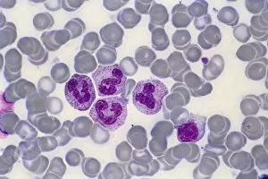
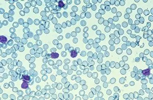
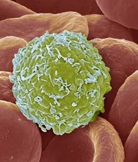
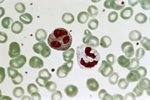
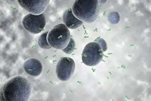
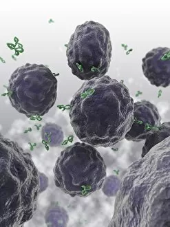
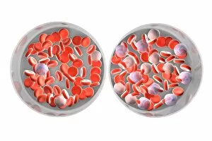
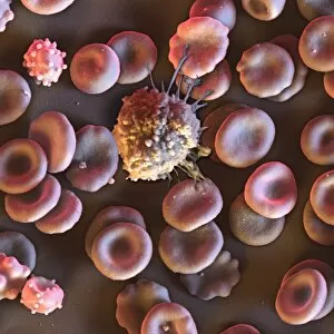
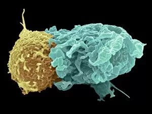
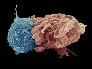
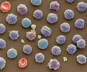
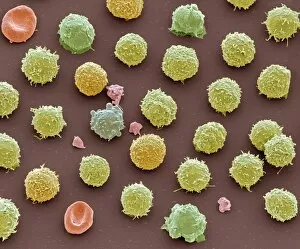
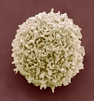
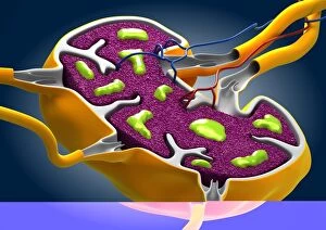
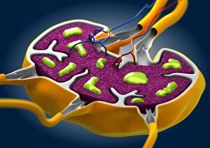
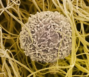
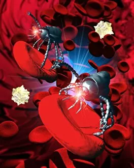
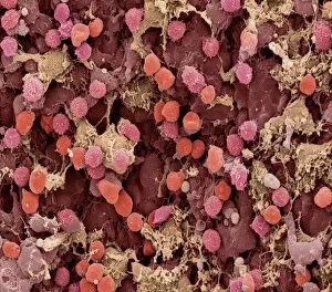
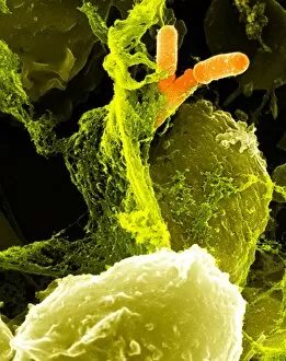
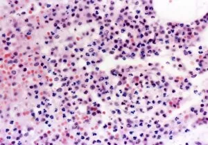
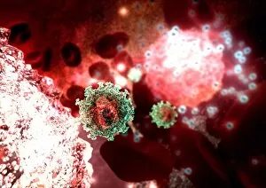
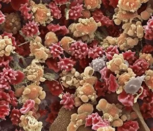
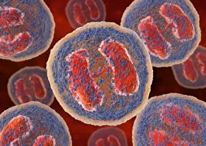
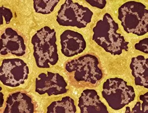
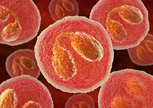
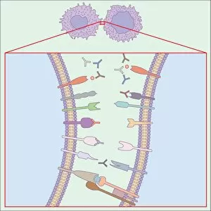
"Exploring the Intricate World of Leucocytes: A Journey through Blood Coagulation Cascade and Cellular Artwork" In this captivating caption, we delve into the fascinating realm of leucocytes, also known as white blood cells. These microscopic warriors play a crucial role in our immune system's defense against harmful invaders. The journey begins with artwork C016/9873, which depicts the intricate process of the blood coagulation cascade. This mesmerizing image showcases how leucocytes work alongside other components to form clots and prevent excessive bleeding. Moving on to scanning electron microscopy (SEM), we witness the beauty of red and white blood cells in stunning detail. SEM images such as C016/3099 and C016/3098 showcase these cellular marvels side by side with platelets, highlighting their collective effort in maintaining our health. Transitioning to transmission electron microscopy (TEM), plasma cells take center stage in TEM imagery. These specialized white blood cells are responsible for producing antibodies that target specific pathogens, safeguarding us from infections. The TEM image reveals their unique structure and function (TEM). Next up is an activated macrophage captured through SEM (C015/6375). This powerful defender engulfs foreign particles or damaged cells, playing a critical role in immune responses. Artwork showcasing lymphocyte white blood cells reminds us of their diversity and importance within our immune system's arsenal. Each type has its own distinct function but works together harmoniously to protect our bodies from harm. As we explore further under SEM lenses, monocyte white blood cell (SEM C016 / 3089) comes into view – another versatile player capable of transforming into different types of immune defenders when needed. No exploration would be complete without acknowledging human red blood cells' essential role in transporting oxygen throughout our bodies efficiently - beautifully showcased via SEM imagery. Finally, multiple SEM images highlight various aspects of general "blood cell" morphology, emphasizing the intricate beauty within our circulatory system.