
Leukocytes Collection (#2)
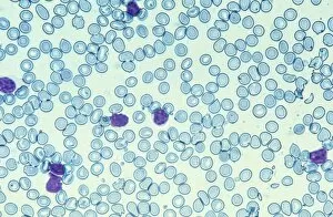
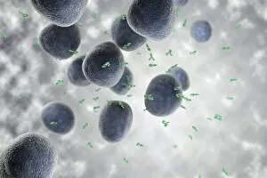
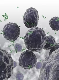
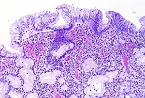
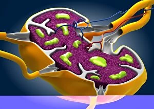
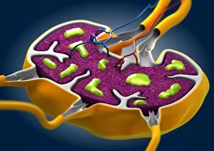
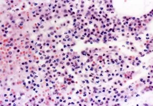
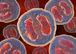
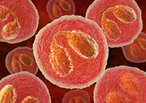
"Exploring the Dynamic World of Leukocytes: Unveiling their Intricate Structures and Functions" Plasma cells, captured under a TEM microscope


For sale as Licensed Images
Choose your image, Select your licence and Download the media
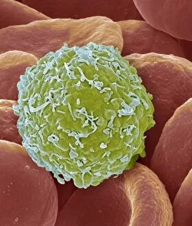
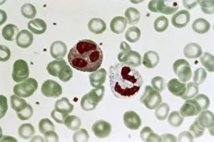
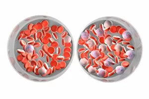
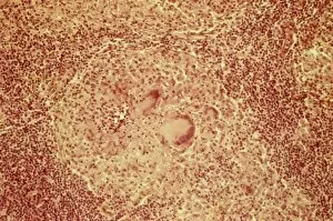
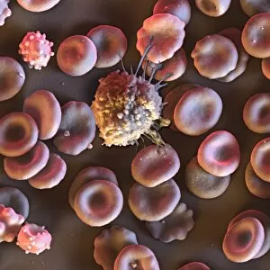
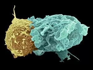
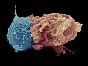
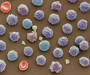
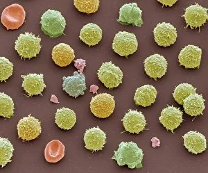
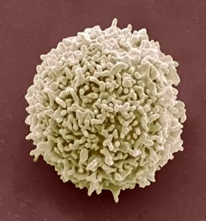
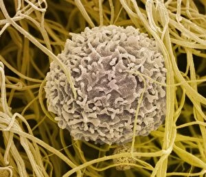
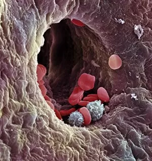
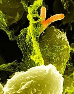
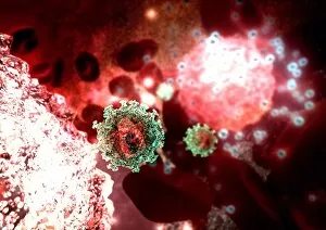
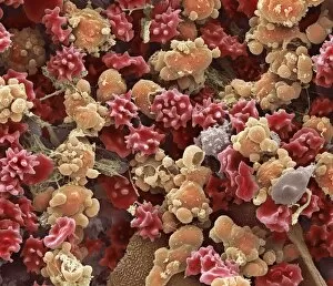
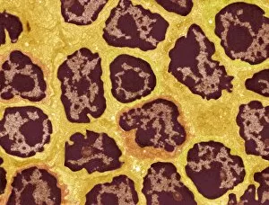
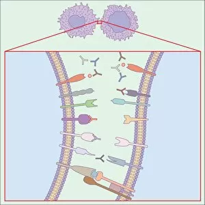
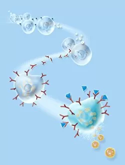
"Exploring the Dynamic World of Leukocytes: Unveiling their Intricate Structures and Functions" Plasma cells, captured under a TEM microscope, reveal their unique morphology and specialized role in producing antibodies to fight off infections. #MicroscopicMarvels Witness the remarkable transformation of an activated macrophage through SEM imaging (C015/6375). These powerful immune cells engulf and destroy harmful pathogens, safeguarding our bodies from harm. #MightyMacrophages Delve into the realm of lymphocyte white blood cells with captivating artwork that showcases their diversity and crucial role in orchestrating our body's immune response against invaders. #GuardiansOfImmunity Behold the intricate network formed by white blood cells and platelets as seen through SEM imaging (C016/3099). This symbiotic relationship ensures efficient clotting mechanisms while defending against foreign substances within our bloodstream. Dive deeper into the mesmerizing world of white blood cells and platelets with another stunning SEM image (C016/3098), revealing their incredible structures up close. #CellularWonders Explore the fascinating features of monocyte white blood cell under SEM microscopy (C016/3089) - these versatile warriors play a vital role in phagocytosis, clearing away debris, and initiating immune responses when needed. Picture No. 11675574 takes us on an extraordinary journey inside our bloodstream, showcasing various types of blood cells captured using high-resolution SEM technology – a testament to nature's beauty at its smallest scale. 8 & 9: Step into a microscopic wonderland where red and white blood cells dance together harmoniously as observed through breathtaking SEM images (#BloodCellsSEM). Marvel at this delicate balance essential for maintaining optimal health. 10: Eosinophil white blood cell steals the spotlight under TEM microscopy (C014/1438).