Antigen presentation, SEM C016 / 3104

![]()
Wall Art and Photo Gifts from Science Photo Library
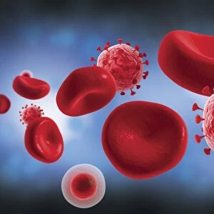
Antigen presentation, SEM C016 / 3104
Antigen presentation. Coloured scanning electron micrograph (SEM) showing the interaction between a macrophage (brown) and a T helper lymphocyte (Th cell, red), two components of the bodys immune system. Both are types of white blood cell. Macrophages are antigen-presenting cells (APCs). They present antigens (fragments on the surface of pathogens or foreign objects) to T lymphocytes, activating them. Each T lymphocyte recognises and binds to a specific antigen. Binding of the Th cell to the antigen presented by the macrophage activates the Th cell. This leads to its proliferation and the activation of other immune cells that eliminate the antigen. Magnification: x7000 when printed 10cm wide
Science Photo Library features Science and Medical images including photos and illustrations
Media ID 9202941
© STEVE GSCHMEISSNER/SCIENCE PHOTO LIBRARY
Activating Activation Adaptive Immunity Antigen Colored Contact Defence Immune Immune Cell Immune System Immunity Immunological Immunology Interacting Interaction Leucocyte Leucocytes Leukocyte Leukocytes Macrophage Monocyte Presentation Presenting Specific Stimulating Stimulation System T Cell White Blood Cell Cells Cutouts
EDITORS COMMENTS
This print titled "Antigen Presentation" offers a stunning glimpse into the intricate world of our body's immune system. In this colored scanning electron micrograph (SEM), we witness the fascinating interaction between a macrophage and a T helper lymphocyte, both essential components of our defense mechanism. The brown-hued macrophage is an antigen-presenting cell (APC) responsible for presenting antigens - fragments found on pathogens or foreign objects - to T lymphocytes. The red-colored T helper lymphocyte, also known as Th cell, plays a crucial role in recognizing and binding to specific antigens presented by the macrophage. As depicted in this image, the binding of the Th cell to the antigen activates it, leading to its proliferation and triggering other immune cells that work together to eliminate the harmful antigen. This process highlights how our immune system responds with precision and specificity when faced with potential threats. With a magnification of x7000 when printed at 10cm wide, this SEM image showcases remarkable detail that allows us to appreciate the complexity and beauty within our own bodies. It serves as a reminder of the incredible biological processes constantly occurring within us, protecting us from harm and maintaining our overall health. Photographed by Steve Gschmeissner from Science Photo Library, this visually striking print combines artistry with scientific exploration, offering viewers an opportunity to marvel at nature's wonders through cutting-edge technology.
MADE IN THE USA
Safe Shipping with 30 Day Money Back Guarantee
FREE PERSONALISATION*
We are proud to offer a range of customisation features including Personalised Captions, Color Filters and Picture Zoom Tools
SECURE PAYMENTS
We happily accept a wide range of payment options so you can pay for the things you need in the way that is most convenient for you
* Options may vary by product and licensing agreement. Zoomed Pictures can be adjusted in the Cart.