
Ligaments Collection (#2)
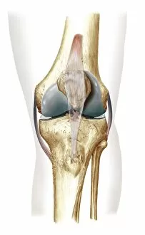
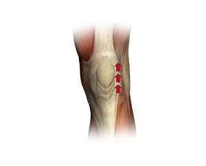
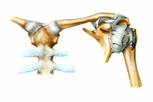
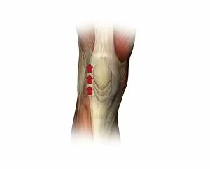
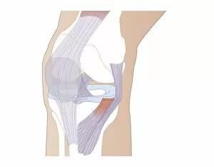
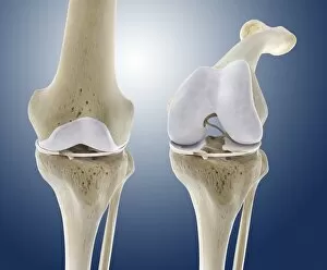
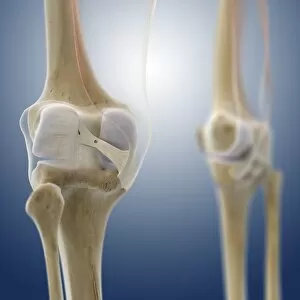
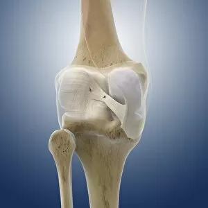
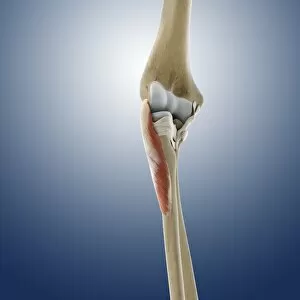
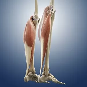
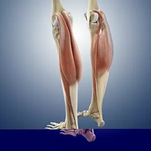
"Ligaments: The Unsung Heroes of Our Body's Stability and Mobility" Damaged knee ligament, artwork: A visual representation of the intricate network in our knees


For sale as Licensed Images
Choose your image, Select your licence and Download the media
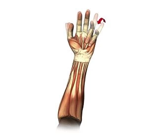
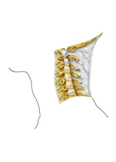
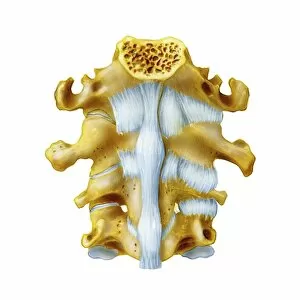
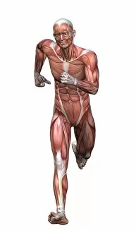
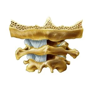
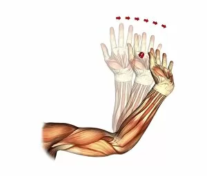
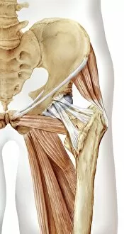
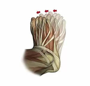
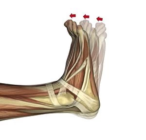
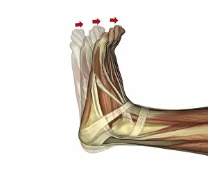
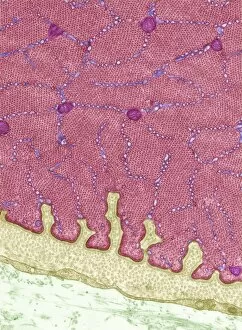
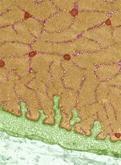
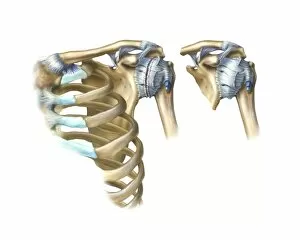
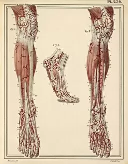
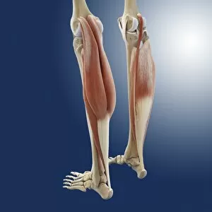
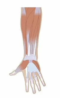
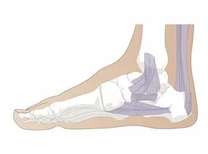
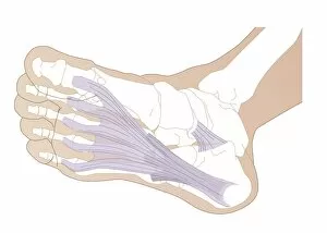
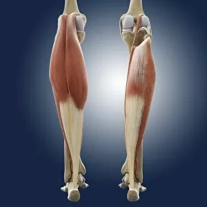
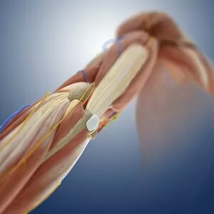
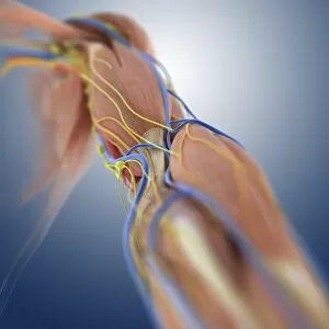
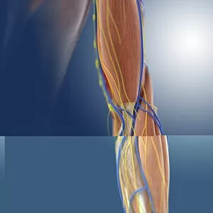
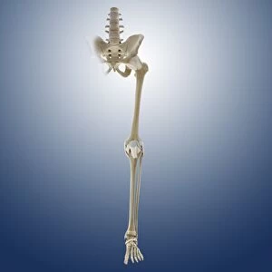
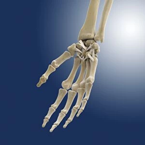
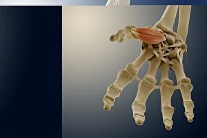
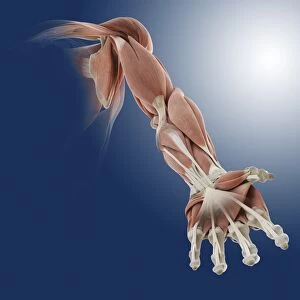
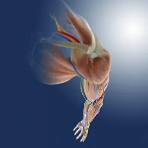
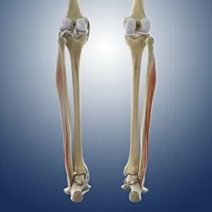
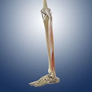
"Ligaments: The Unsung Heroes of Our Body's Stability and Mobility" Damaged knee ligament, artwork: A visual representation of the intricate network in our knees, highlighting the importance of their role in supporting joint stability. Skeleton & Ligaments: Explore the hidden framework beneath our skin as we delve into the fascinating connection between our skeleton and ligaments that enable us to move with grace. Arm circulation, anatomical artwork C013 / 7419: Discover how ligaments work hand-in-hand with blood vessels to ensure proper arm circulation, allowing us to perform everyday tasks effortlessly. Muscles of the neck: Unveil the interplay between muscles and ligaments in our neck region, crucial for maintaining posture and facilitating smooth head movements. Eye muscle, TEM C014 / 1468: Delicate yet powerful eye muscles intricately intertwined with resilient ligaments—uncover this captivating combination responsible for precise eye movements. Outer ankle ligaments, artwork C013 / 4452: Step into a world where outer ankle ligaments provide stability during every stride we take—a testament to their resilience against external forces. Inner ankle ligaments, artwork C013 / 4451: Journey deeper into understanding inner ankle ligament anatomy as they safeguard against inward rolling motions while walking or running. Eye anatomy, SEM: Peer through a scanning electron microscope lens at detailed images revealing how tiny but mighty eye structures rely on robustly connected networks of supportive ligaments. Muscles (engraving): Witness an enchanting engraving showcasing various muscles working harmoniously alongside essential connective tissues like tendons and—of course—ligaments. Ligaments of the human body (engraving): An artistic masterpiece capturing an array of interconnected human body parts held together by strong bands known as "ligaments"—a true testament to the body's architectural brilliance.